Angiographic method and apparatus allowing identification of contrast agent propagation

a technology of contrast agent and detection method, applied in the field of angiographic method and apparatus allowing identification of contrast agent propagation, can solve problems such as time saving

Inactive Publication Date: 2012-02-14
SIEMENS HEALTHCARE GMBH
View PDF6 Cites 6 Cited by
  • Summary
  • Abstract
  • Description
  • Claims
  • Application Information

AI Technical Summary

Benefits of technology

[0008]Such a method for acquisition of angiographic data sets has a number of advantages. First, the method can be implemented without manual intervention of a user, which leads to a time savings. Furthermore, with the method it is possible to determine the arrival of the second propagation phase of the contrast agent. An image reconstruction can thereby ensue in a targeted manner using only the data sets that were acquired during the first propagation phase of the contrast agent. For example, an image reconstruction can be implemented from the magnetic resonance data sets to generate an arteriograph. Data sets that were acquired during the second propagation phase (for example the venous phase) are then not taken into account, and thus are only shown in the reconstructed image data. It is thus no longer necessary to manually differentiate between arteries and veins in the image data. Such an automatic determination of the arrival of the second propagation also possesses the advantage that an incorrect association of data sets with the first or second propagation phase is avoided. For example, the times for a propagation of the contrast agent in the different phases for different examined persons can deviate significantly from one another depending on the circulatory situation, and thus an automatic determination of the arrival of the second propagation phase is particularly advantageous.
[0018]Furthermore, a magnetic resonance system is provided for acquisition of angiographic magnetic resonance data sets of a region of an examination subject to be examined in whose bloodstream a contrast agent has been introduced, wherein the magnetic resonance system possesses: an acquisition unit that acquires MR data sets from the region to be examined; a control unit that controls the acquisition of MR data sets such that an MR data set of the region to be examined is acquired as a reference data set during a first propagation phase of the contrast agent in the bloodstream, and the additional MR data sets of the region to be examined are acquired; and a computer that is designed such that it executes the following steps: determination of reference data from the reference data set; determination, on the basis of the reference data, of variations that are caused in at least some of the additional MR data sets due to an arrival of a second propagation phase of the contrast agent; and determination of the arrival of the second propagation phase of the contrast agent in the bloodstream on the basis of the determined variations. The computer advantageously implements these steps automatically. Such a magnetic resonance system enables the arrival of the second propagation phase to be automatically determined. This enables a reconstruction of image data to be implemented based on the magnetic resonance data sets that were acquired during the first propagation phase. The magnetic resonance system can furthermore be designed to implement one of the methods described above.

Problems solved by technology

First, the method can be implemented without manual intervention of a user, which leads to a time savings.

Method used


Image

  • Angiographic method and apparatus allowing identification of contrast agent propagation
  • Angiographic method and apparatus allowing identification of contrast agent propagation
  • Angiographic method and apparatus allowing identification of contrast agent propagation

Examples


Embodiment Construction

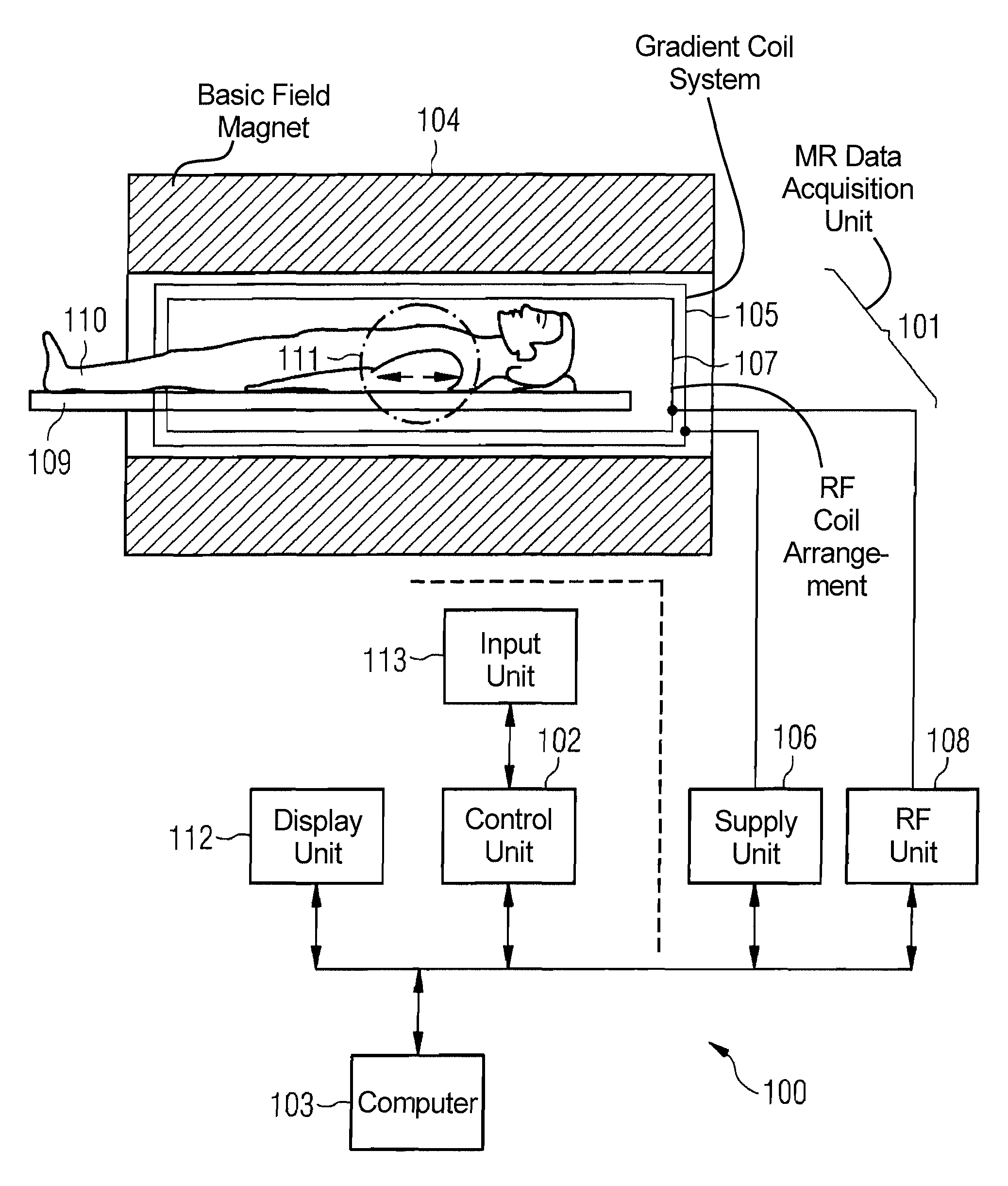
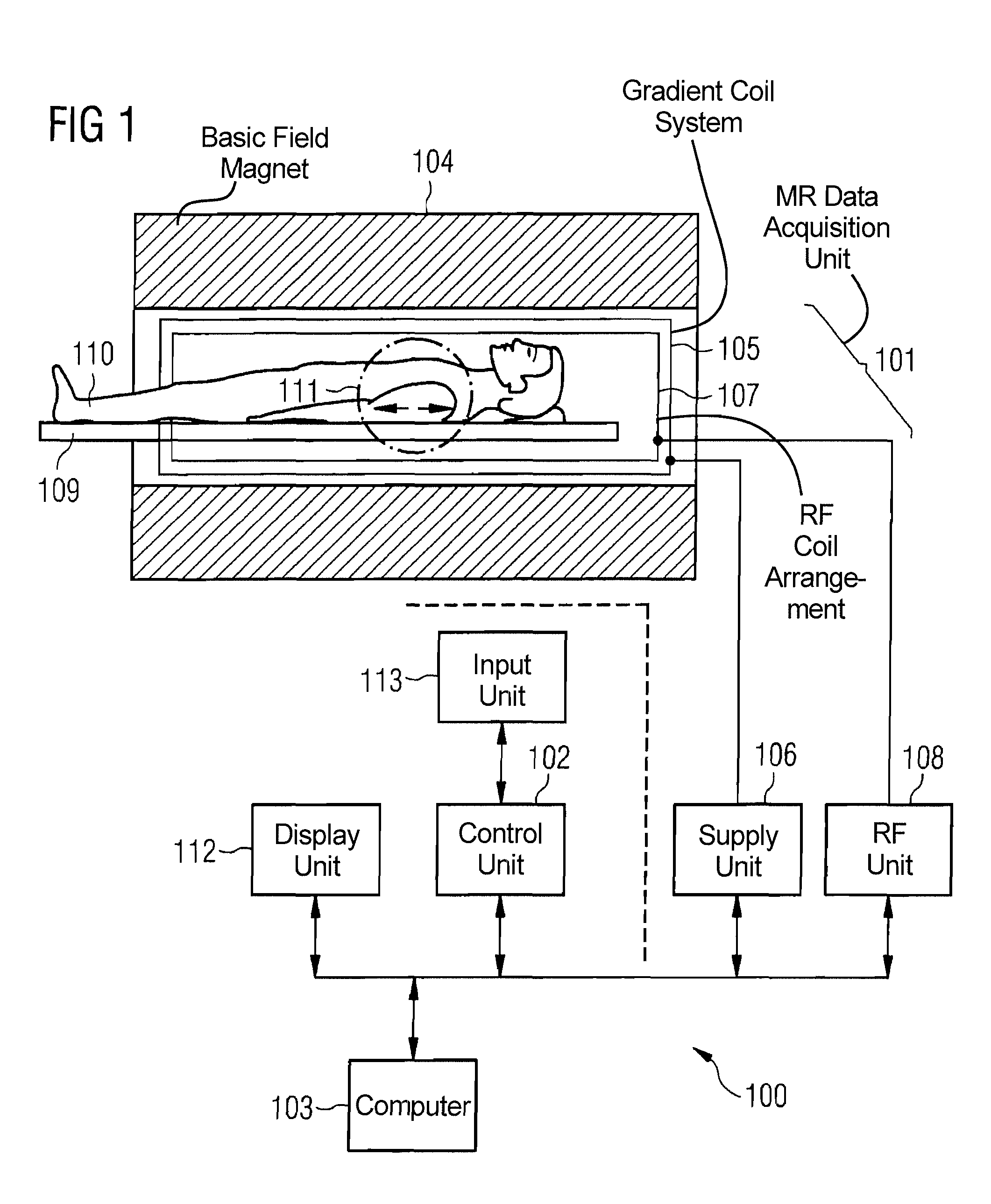
[0025]FIG. 1 schematically shows a magnetic resonance system 100. The magnetic resonance system 100 has an MR acquisition unit 101 to acquire magnetic resonance signals (magnetic resonance data sets) as well as a control unit 102 and a computer 103. The acquisition unit 101 has the components that are necessary to acquire magnetic resonance signals. Among these are, for example, a basic field magnet 104 that generates a polarization field B0, a gradient coil system 105 and a supply unit 106 to generate and apply magnetic field gradients, for example a slice selection gradient, a phase coding gradient or a frequency coding gradient that are used for an imaging and spatial coding, as well as a radio-frequency (RF) coil arrangement 107 to radiate RF pulses and an induction coil to acquire magnetic resonance signals. Furthermore, the magnetic resonance system can possess a movable examination table 109 on which, for example, an examined person 110 is placed to traverse the magnet 104 of...


PUM


Abstract

In a method and apparatus for acquisition of angiographic data sets of a region of an examination subject, into whose bloodstream a contrast agent has been introduced, a data set of the region to be examined is acquired during a first propagation phase of the contrast agent in the bloodstream as a reference data set to determine reference data, additional data sets of the region to be examined are then acquired, and based on the reference data, variations that are caused in at least some of the additional data sets due to the arrival of a second propagation phase of the contrast agent are determined, and the arrival of the second propagation phase of the contrast agent in the bloodstream is identified based on the determined variations.

Description

BACKGROUND OF THE INVENTION[0001]1. Field of the Invention[0002]The present invention concerns a method for acquisition of angiographic data sets from a region of an examination subject, as well as a magnetic resonance system for acquisition of such data sets.[0003]2. Description of the Prior Art[0004]Angiography is a known medical diagnosis method in which blood vessels are shown by means of diagnostic imaging methods. For example, x-ray or magnetic resonance tomography methods can be used for this purpose. In magnetic resonance (MR) tomography, for example, the imaging is sensitive to the movement of nuclear spins that generate the MR signal. These effects can be used to acquire angiographic images, i.e. images of the vessels of the examined person. Angiographic techniques that are based only on the flux effects of the spins located in the body are known for this. Furthermore, contrast agent-supported MR angiography (ceMRA) is a method frequently applied in clinical practice. With...

Claims


Application Information

Patent Timeline
Patent Type & Authority Patents(United States)
IPC IPC(8): A61B5/055A61B6/00G01V3/00
CPCA61B5/055A61B5/7289G01R33/5601G01R33/5635G01R33/56509
Inventor SCHMIDT, SEBASTIAN
Owner SIEMENS HEALTHCARE GMBH
Why Eureka
Try Eureka