
Human intestinal cancer primary cells, applications and culture methods
A primary cell and intestinal cancer technology, applied in cell culture active agents, tumor/cancer cells, biochemical equipment and methods, etc., can solve the problems of poor cell state, culture failure and death of primary human intestinal cancer cells
Patent Information
- Authority / Receiving Office
- CN · China
- Patent Type
- Patents(China)
- Current Assignee / Owner
- Publication Date
- 2021-08-17
Smart Images
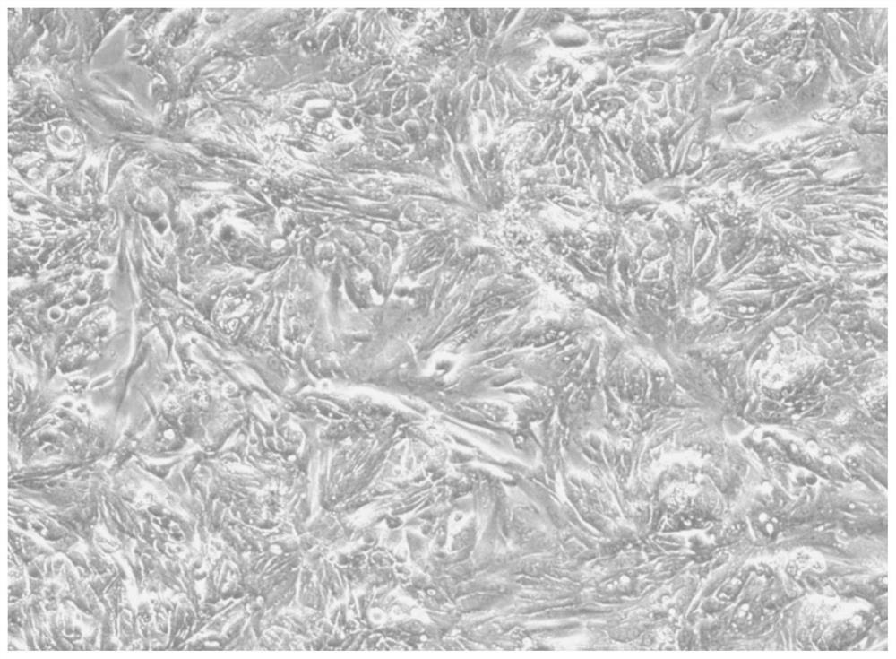
Figure 1 
Figure 2 
Figure 3
Abstract
Description
technical field
[0001] The invention relates to the field of biotechnology, in particular to primary human intestinal cancer cells, application and culture method. Background technique
[0002] Intestinal cancer is a common digestive tract disease. Generally, the biological characteristics of primary cultured tumor cells have not changed much because the tissue has just been isolated from the body, and the original genetic characteristics are still retained. The gene retention is more than 90%. It is suitable for biochemical molecular Experimental and drug susceptibility tests and related experiments to explore the mechanism, the data are more convincing. The establishment of primary human intestinal cancer cells is of great significance for the establishment of cell lines, drug sensitivity testing experiments, personalized treatment, and research on the pathogenesis and therapeutic mechanism of intestinal cancer.
[0003] At present, primary human intestinal cancer cells a...
Examples
Embodiment 1
[0075] Example 1: Isolation and culture of human intestinal cancer primary cells
[0076] (1) With the informed consent of the patient or the patient's guardian, collect part of the intestinal cancer tissue removed during the patient's operation;
[0077] (2) Transfer the intestinal cancer tissue collected in step (1) to a cell culture dish, wash with a cleaning solution (containing 0.4 mg / ml penicillin, 0.4 mg / ml kanamycin sulfate, 0.4 μg / ml amphotericin B, 4 μg / ml vancomycin in normal saline) for 8 times to remove non-cancerous tissue impurities such as fat and mucous membrane;
[0078] (3) Transfer the cleaned intestinal cancer tissue in step (2) to a new culture dish, cut the cleaned intestinal cancer tissue into small pieces with scissors and a blade, and chop them into fine pieces;
[0079] (4) Transfer the crushed intestinal cancer tissue to a 50ml centrifuge tube, centrifuge at 2000rpm for 8min, remove the supernatant, add 9ml DF medium for resuspension, and then add ...
Embodiment 2
[0084] Example 2: Purification of Human Intestinal Cancer Primary Cells
[0085] (1) When the confluence of the cells cultured in step (8) of Example 1 reaches 70-90%, the serum-free medium is removed and digested with 1-2 ml EDTA-Trypsin.
[0086] (2) Observe under a microscope until the cells are detached from the culture flask, discard the fibroblasts digested by EDTA-Trypsin first, and then collect the digested cells while digesting, and stop the digestion of the collected cells with DF10 medium until All cells were digested to completion.
[0087] (3) Centrifuge at 1500rpm for 6min, remove the supernatant, resuspend with serum-free medium, and store at 37°C, 5% CO 2Place in the cell incubator for 1.5 hours, after the fibroblasts adhere to the wall, collect the unattached cells, transfer them to a new culture bottle to continue culturing, and repeat the adhesion for 3 times to achieve the effect of completely removing the fibroblasts. The primary human intestinal cancer ...
Embodiment 3
[0089] Embodiment 3: Morphological observation
[0090] By observing under an inverted microscope, it can be found that the human intestinal cancer primary cells cultured after Examples 1 and 2 are in the form of epithelial cells. Cells proliferate rapidly, without contact inhibition, squeeze each other, and appear stacked or mosaic. For specific results, see figure 1 .