
Microscope and method for light field microscopy and confocal microscopy with light sheet excitation
A technology of microscopes and light sources, applied in the field of microscopes, can solve problems such as equipment production cost constraints
Patent Information
- Authority / Receiving Office
- CN · China
- Current Assignee / Owner
- Publication Date
- 2022-05-06
Smart Images
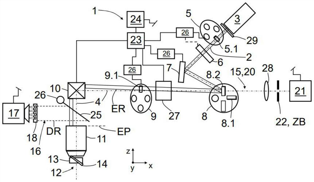
Figure 1 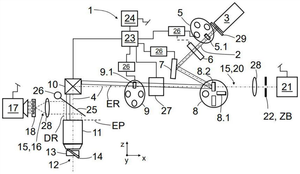
Figure 2 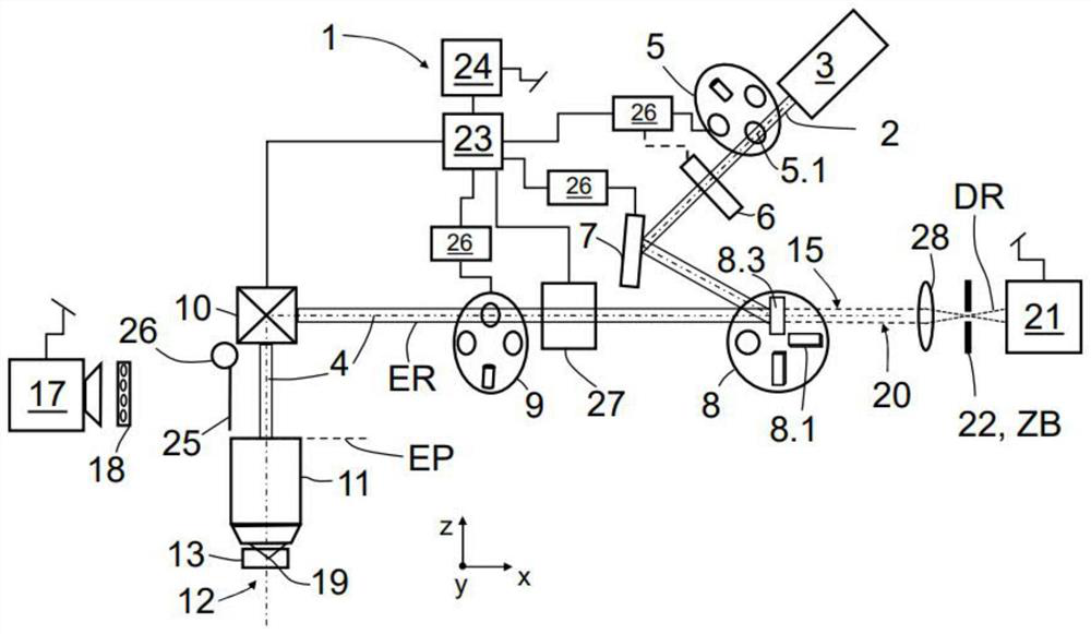
Figure 3
Abstract
Description
technical field
[0001] The present invention relates to a microscope and to a method for operating a microscope for light field microscopy and confocal microscopy with light sheet excitation. Background technique
[0002] Modern microscopy increasingly focuses on the rapid capture of three-dimensional sample volumes. An important application in this context is the measurement of neural signals in networks of neural cells. These networks are spread across the brain over hundreds or thousands of microns. To be able to understand the fundamental capabilities of the brain, the responses of the network or large parts thereof need to be captured as completely as possible with high temporal resolution. Here, as it is important to understand not only morphological structures but also functional processes, these approaches are also combined under key functional imaging.
[0003] In the field of microscopy, many different methods are known with which functional imaging can be appro...
Examples
Embodiment Construction
[0083] exist figure 1 The basic setup of the microscope 1 according to the invention is schematically shown in . For the sake of clarity, the description only shows technical elements necessary to explain the present invention. The light source 3 is arranged in the excitation beam path 2 of the microscope 1 and is embodied in particular in the form of a laser light source 3 which emits excitation radiation ER along the optical axis 4 of the excitation beam path 2 . In the shown first operating state of the microscope 1 (the microscope 1 is used in the first operating mode), there is a first device 5 for introducing an optical element into the excitation beam path 2 and a first cylindrical lens in the form of a first A cylindrical optical element 5.1 is introduced into the path 2 of the excitation beam. Due to the action of the first cylindrical optical element 5 . 1 , the excitation radiation ER is shaped by limiting its cross section transverse to its direction of propagati...