
Method for controlling emmission in an x-ray imaging device
a technology of x-ray imaging and x-ray, which is applied in the field of controlling the x-ray emission of an x-ray imaging device, can solve the problems of inability to adapt, lack of precision, and inability to meet the needs of patients, and achieve the effect of minimizing the x-ray dos
Patent Information
- Authority / Receiving Office
- US · United States
- Patent Type
- Applications(United States)
- Current Assignee / Owner
- Publication Date
- 2013-01-17
Smart Images
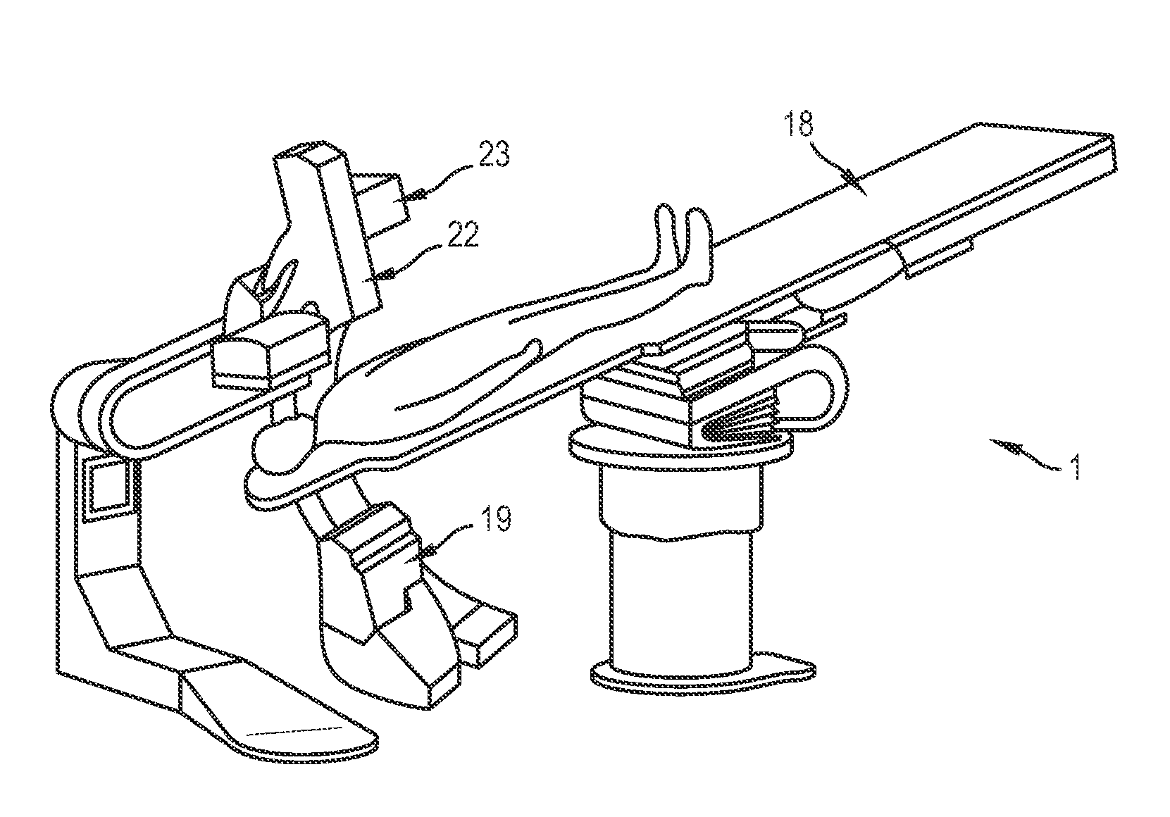
Figure 1 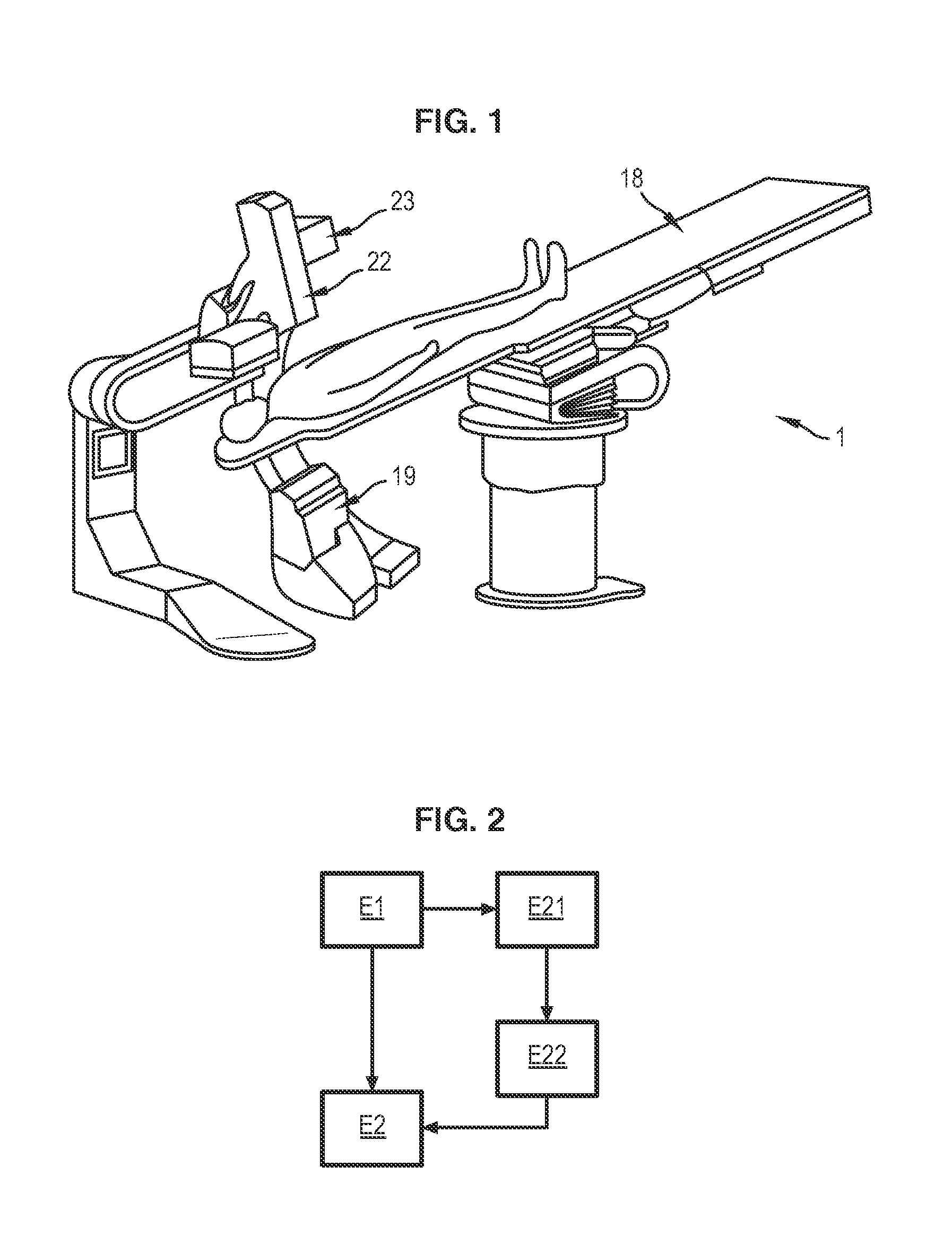
Figure 2 
Figure 3
Abstract
Description
BACKGROUND OF THE INVENTION
[0001] 1. Field of the Invention
[0002] Embodiments of the present invention relate to a method for controlling the X-ray emission of an X-ray imaging device, and an X-ray imaging device capable of implementing the method.
[0003] 2. Description of the Prior Art
[0004] X-ray imaging devices are used in numerous medical applications. More particularly, these devices are used in interventional radiography, which allows a practitioner to monitor the conducting of a procedure being performed on a patient. Interventional radiology is notably used in vascular surgery in neurology and cancerology.
[0005] In these devices, an emitter emits X-rays towards the patient's body, said X-rays then being collected by a detector to allow an image of the patient's body to be obtained. Health regulations require the limiting of unnecessary exposure of patients to X-rays which, at high dose, are harmful for the body.
[0006] Optimization of the X-ray dose emitted by the device includes co...
Examples
Embodiment Construction
[0018]FIG. 1 illustrates an X-ray imaging device 1 according to an embodiment of the present invention. The device is adapted to take images of a patient's body. As is conventional in the art, the device 1 comprises a table 18 on which a patient is positioned. Generally, the patient lies down on the table 18 when images are taken by the device 1.
[0019]The device 1 comprises an X-ray emitter 19 capable of emitting X-rays towards the patient's body. In general, the emitter 19 comprises a vacuum tube comprising an anode and a cathode, in which a filament, typically a coiled tungsten wire, is heated to high temperature by means of an electric current. The beam of electrons generated by the cathode is accelerated towards the anode. The interaction between the incident beam of electrons and the material of the anode allows the generation of X-rays.
[0020]The X-rays pass through the patient's body and are collected by a detector 22 allowing an image of the patient's body to be obtained.
[002...