
Markers of tissue aging and uses therefor
a tissue aging and marker technology, applied in the field of tissue aging markers, can solve the problems of affecting tissue functionality, contributing to aging, damage to proteins, lipids and nucleic acids, etc., and achieve the effects of preventing or reducing the risk of post-dated pregnancy, and reducing the risk of aging or oxidative damag
Patent Information
- Authority / Receiving Office
- US · United States
- Patent Type
- Applications(United States)
- Current Assignee / Owner
- Publication Date
- 2020-10-01
Smart Images
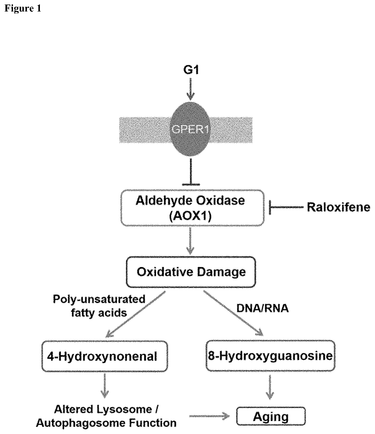
Figure 1 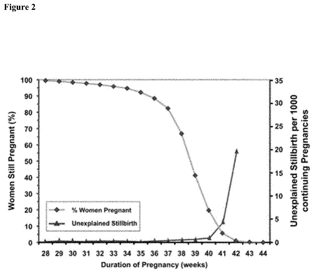
Figure 2 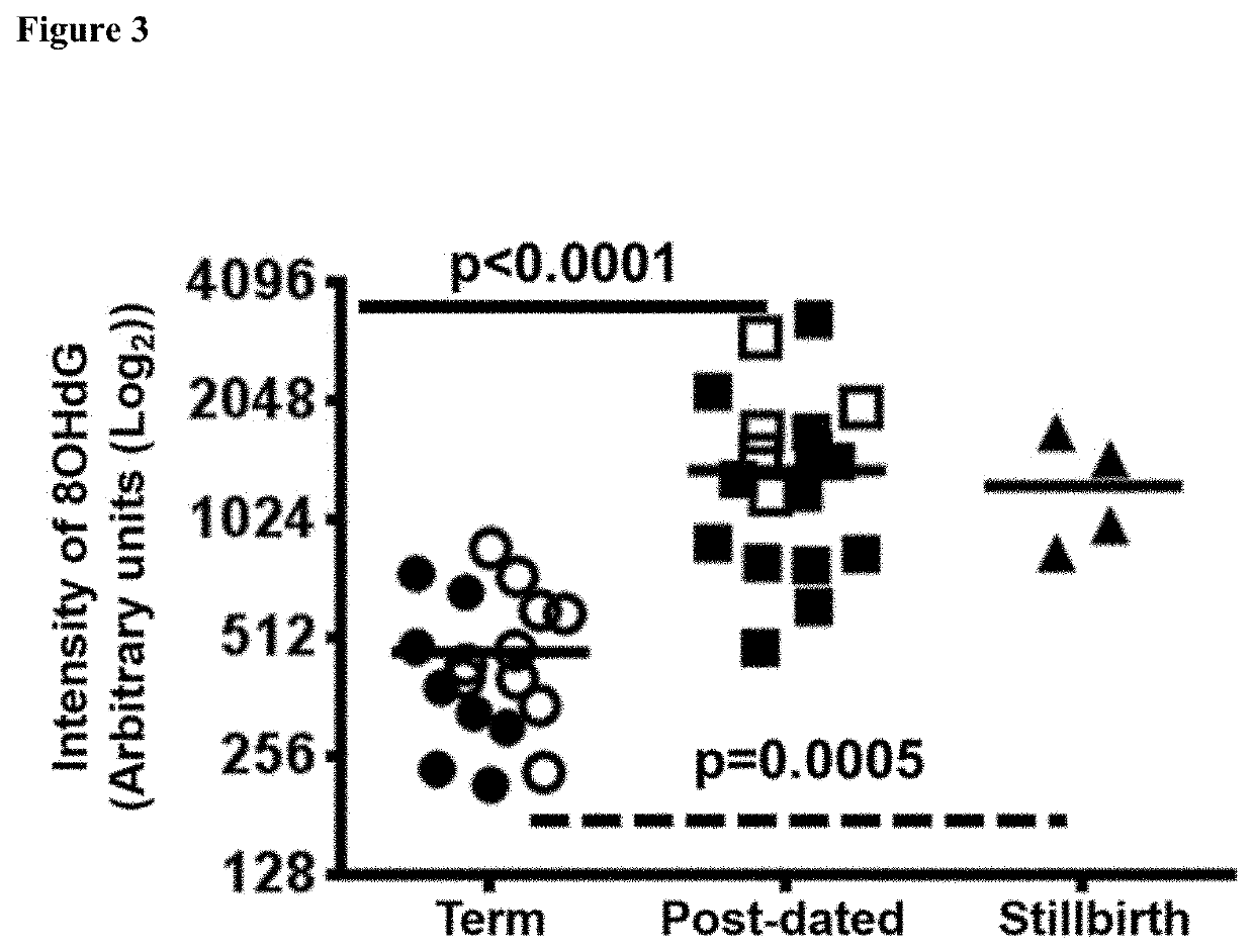
Figure 3
Abstract
Description
CROSS-REFERENCE TO RELATED APPLICATION(S)
[0001] The present application is a U.S. National Stage of International Patent Application No. PCT / AU2018 / 050190, filed on Mar. 2, 2018, and which claims priority to Australian Patent Application No. 2017900740, filed Mar. 3, 2017, both disclosures of which are hereby incorporated by reference in their entirety for all purposes.FIELD OF THE ART
[0002] The present disclosure relates generally to methods of detecting or determining aging and / or oxidative damage in tissue, such as placental tissue, skin, kidney and brain tissue. The disclosure also relates to methods of delaying, slowing or preventing aging and / or oxidative damage in such tissue.SEQUENCE LISTING
[0003] This disclosure, in accordance with 37 CFR § 1.52, incorporates by reference the sequence listing material contained within text file titled “068612.00009_ST25.txt”, created on Jan. 2, 2020 and totaling 7,124 bytes.BACKGROUND
[0004] Accumulated random damage to tissue may affect tissue ...
Examples
example 1
hip Between Stillbirth Risk and Length of Gestation
[0108]To illustrate the relationship between stillbirth risk and length of gestation the inventors created a Kaplan Myer plot of the data on human gestational length in a population with relatively low levels of medical intervention from Omigbodun and Adewuyi (1997, J Natl Med Assoc, 89: 617) and combined it with the data on risk of stillbirth per 1000 continuing pregnancies from Sutan et al. (2010, J Perinatol, 30: 311-318) (FIG. 2). The data illustrate that stillbirth is consistent with an aging aetiology as defined by Johnson et al. (1999, Cell, 96: 291-302).
example 2
nd Lipid Oxidation in Placental Tissue
[0109]As oxidative damage has been observed in many aging tissues, the inventors sought evidence of placental DNA / RNA oxidation as measured by 8-hydroxy-deoxyguanosine / 8-hydroxyguanosine (8OHdG / 8HOG), as a marker of DNA / RNA oxidation. Immunohistochemistry (IHC) was performed in placentas for 8OHdG / 8HOG and the average intensity of 8OHdG / 8HOG staining in nuclei / frame demonstrated a significant increase in DNA / RNA oxidation in post-dated and stillbirth associated placentas (FIG. 3).
[0110]The increase in DNA oxidation suggested free radical damage that might also lead to lipid peroxidation. Lipid peroxidation has been observed to increase in Alzheimer' s disease as measured by the formation of 4-hydroxynonenal (4HNE) (Markesbery and Lovell, 1998, Neurobiol Aging, 19: 33-36). The inventors therefore performed immunohistochemistry for 4HNE in post-dated, stillbirth and term placental tissue. This revealed a marked increase in 4HNE staining in post-da...
example 3
and Clustering of Lysosomes in Post-Dated and Stillbirth Placentas
[0112]Misfolded proteins and damaged mitochondria are normally recycled in autophagosomes in a process that involves autophagosome fusion with proteolytic enzyme containing lysosomes. Accumulation of abnormal protein is thought to play a role in aging particularly in the brain, for instance the accumulation of tau and amyloid protein in Alzheimer's disease and mutant huntin in Huntington's disease. In Huntington's disease, the distribution of the lysosomes within neurones is altered with increased perinuclear accumulation of lysosomes. The inventors used a lysosomal marker, lysosome-associated membrane protein-2 (LAMP2) to analyse the distribution of lysosomes in the placenta by immunohistochemistry. This showed lysosomes positioned on the apical surface of term placental syncytiotrophoblast (FIGS. 6A, 6D and 6E), whereas lysosomes relocated to the perinuclear and the basal surface in post-dated and stillbirth placent...