
Prevention and Treatment of Viral Infection and Viral Infection-Induced Organ Failure
a technology of organ failure and viral infection, applied in the field of compositions, can solve the problems of organ failure, multiple organ failure, known cause of mortality, etc., and achieve the effects of reducing the mortality rate, and mitigating the fibrosis of one or more organs
Patent Information
- Authority / Receiving Office
- US · United States
- Patent Type
- Applications(United States)
- Current Assignee / Owner
- Publication Date
- 2021-10-14
Smart Images
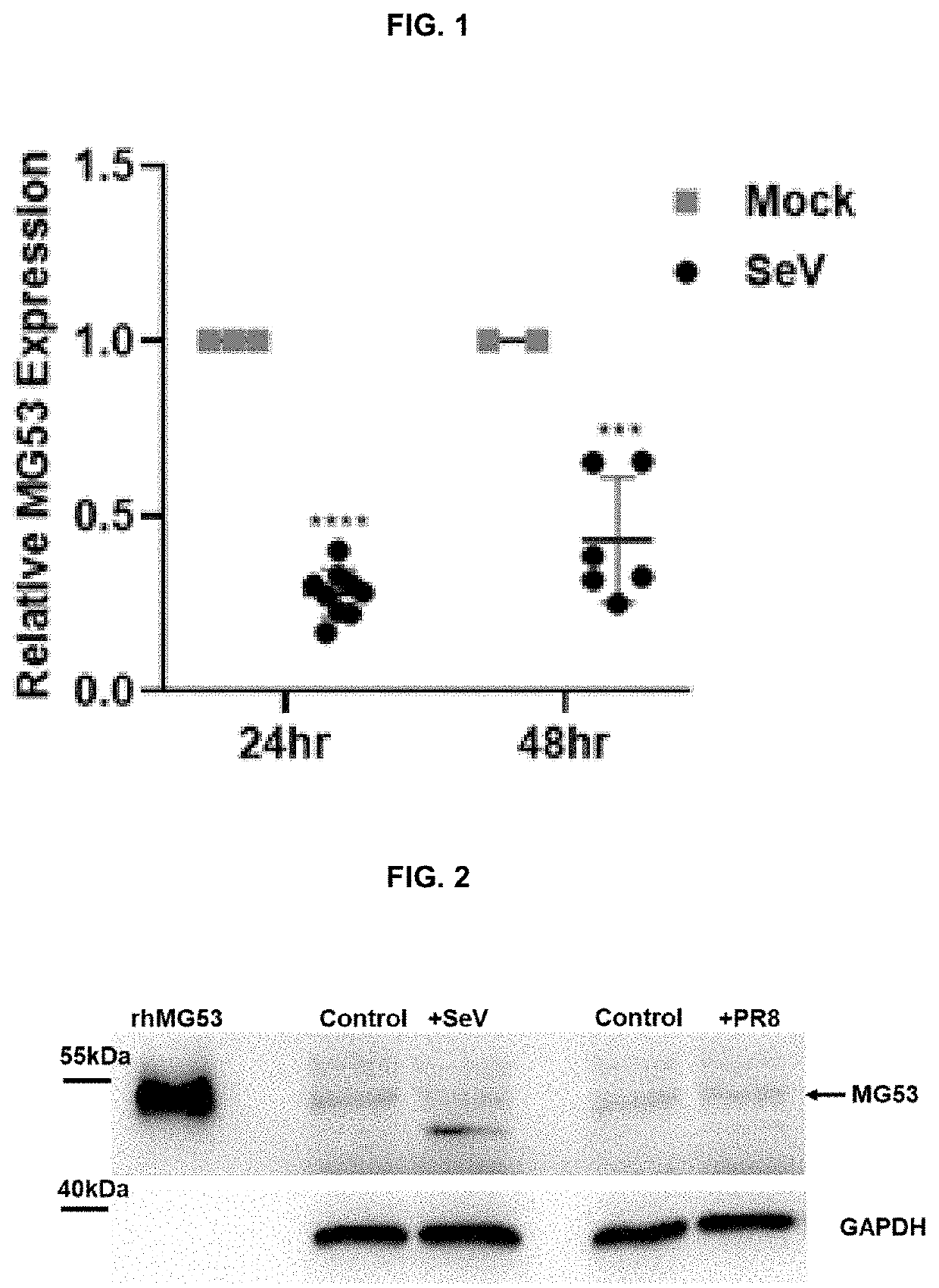
Figure 1 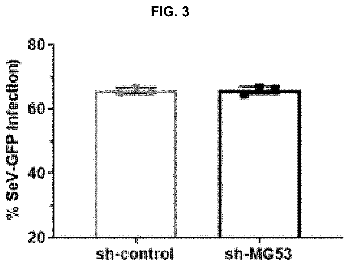
Figure 2 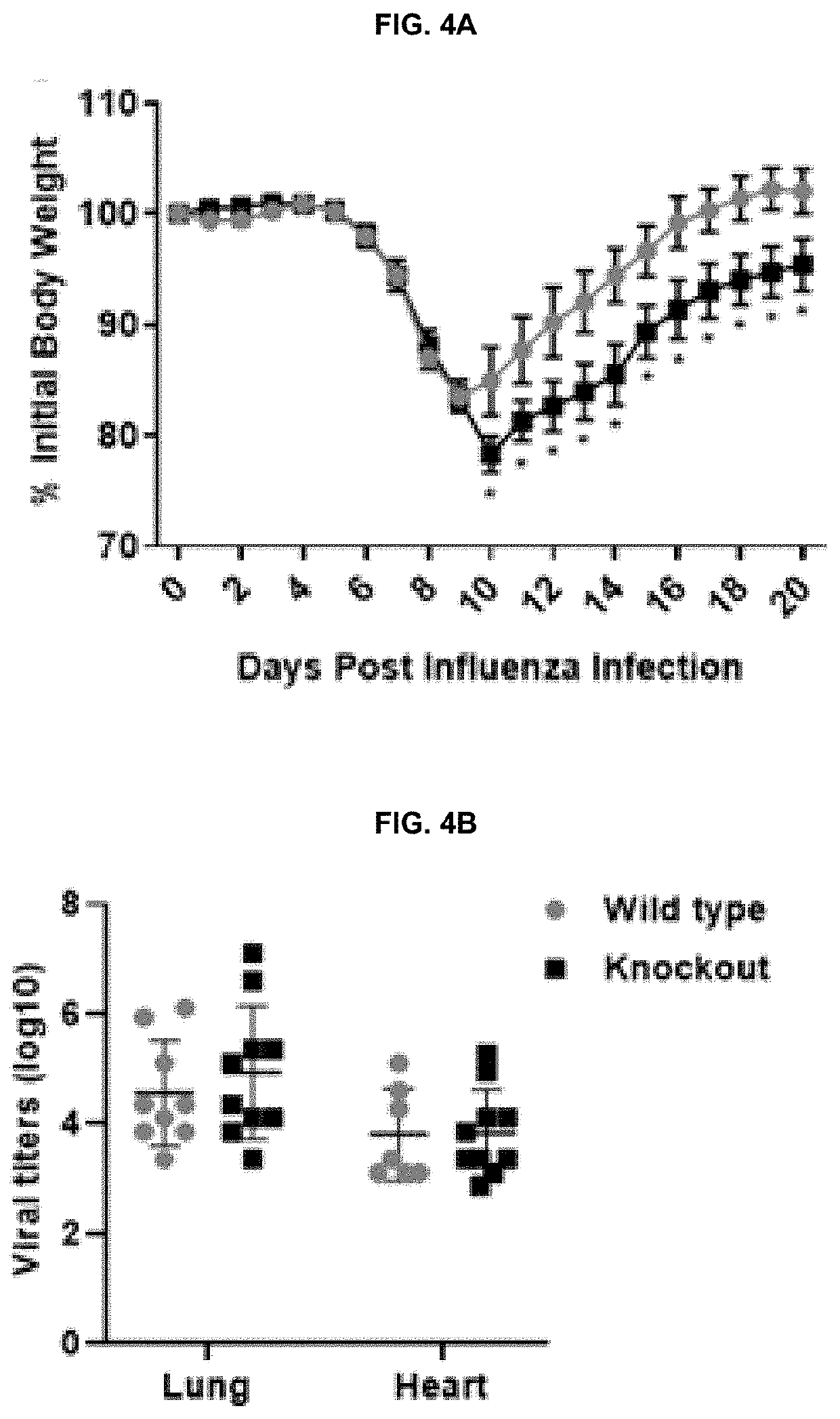
Figure 3
Abstract
Description
CROSS-REFERENCE TO EARLIER FILED APPLICATION
[0001] This application claims the benefit of and is a continuation-in-part of PCT / US20 / 28112 filed Apr. 14, 2020, the entire disclosure of which is hereby incorporated by reference.STATEMENT REGARDING FEDERALLY SPONSORED RESEARCH
[0002] The U.S. Government has certain rights in this invention pursuant to the following grants. This work was supported by grants from the National Institutes of Health (NIH) to Dr. Jianjie Ma (grants No. DK106394, No. AR061385, No. AR070752, and No. HL138570), Department of Defense grant to Dr. Jianjie Ma (DoD PR170989), and NIH grants to Dr. Jacob S. Yount (grants No. AI130110 and No. AI142256). This work was also supported by a NIH T32 fellowship to Adam D. Kenney (funded by grant No. GM068412).INCORPORATION BY REFERENCE
[0003] In compliance with 37 CFR 1.52(e)(5), the instant application contains Sequence Listings which have been submitted in electronic format via EFS and which are hereby incorporated by referen...
Examples
example 1
In Vitro Assay in THP1 Cells: SeV
[0136]Sendai virus (SeV) expressing GFP, SeV strain Cantell, PR8 were propagated in embryonated chicken eggs and titered on LLCMK2 cells for SeV and MDCK cells for influenza virus. SeV-GFP and SeV infections were allowed to proceed for 24 or 48 hours using multiplicity of infections (MOIs) of 2 and 5 respectively. 24 hrs post SeV-GFP infection, THP1 cells were washed in PBS and fixed using 4% paraformaldehyde. Cells were washed, resuspended in PBS, and analyzed with a FACSCanto II flow cytometer (BD Biosciences) to determine the percentage of GFP positive cells. Data was analyzed using FlowJo software.
example 2
In Vitro Assay in THP1 Cells: SeV and H1N1 Influenza
[0137]Sendai virus (SeV) expressing GFP, SeV strain Cantell, and influenza virus strain PR8 were propagated in embryonated chicken eggs and titered on LLCMK2 cells for SeV and MDCK cells for influenza virus. SeV-GFP and SeV infections were allowed to proceed for 24 or 48 hours using multiplicity of infections (MOIs) of 2 and 5 respectively. 24 hrs post SeV-GFP infection, THP1 cells were washed in PBS and fixed using 4% paraformaldehyde. Cells were washed, resuspended in PBS, and analyzed with a FACSCanto II flow cytometer (BD Biosciences) to determine the percentage of GFP positive cells. Data was analyzed using FlowJo software.
example 3
Knockdown of MG53 in THP1 Cells
[0138]Control shRNA (SEQ ID 1: 5′-GACTGACATGTCAAGCTGTAC-3′) and MG53 shRNA (SEQ ID 2: 5′-GAAGAGTGTGGCTGTGCTGGAGCATCAGC-3′) were ligated into pKLO-mcherry-puro vector. In brief, HEK293-FT cells were transfected with packaging, envelope, and target plasmids. Media was changed 18 hours after transfection, followed by collection of virus-containing media 48 hours later. Virus-containing media was centrifuged at 1200×g for 5 min and filtered with 0.45 μm filters. THP1 cells were then incubated with viral media. After 24 hrs, media was replaced, and cells were allowed 48 hrs to recover. Following recovery, cells were selected for using puromycin (1.0 μg / mL), and subsequently cultured in RPMI-1640 media supplemented with puromycin (0.5 μg / mL), to generate sh-control and sh-MG53 THP1 cells.