
Low cost fundus imager with integrated pupil camera for alignment aid
Patent Information
- Authority / Receiving Office
- US · United States
- Current Assignee / Owner
- Publication Date
- 2021-12-02
Smart Images
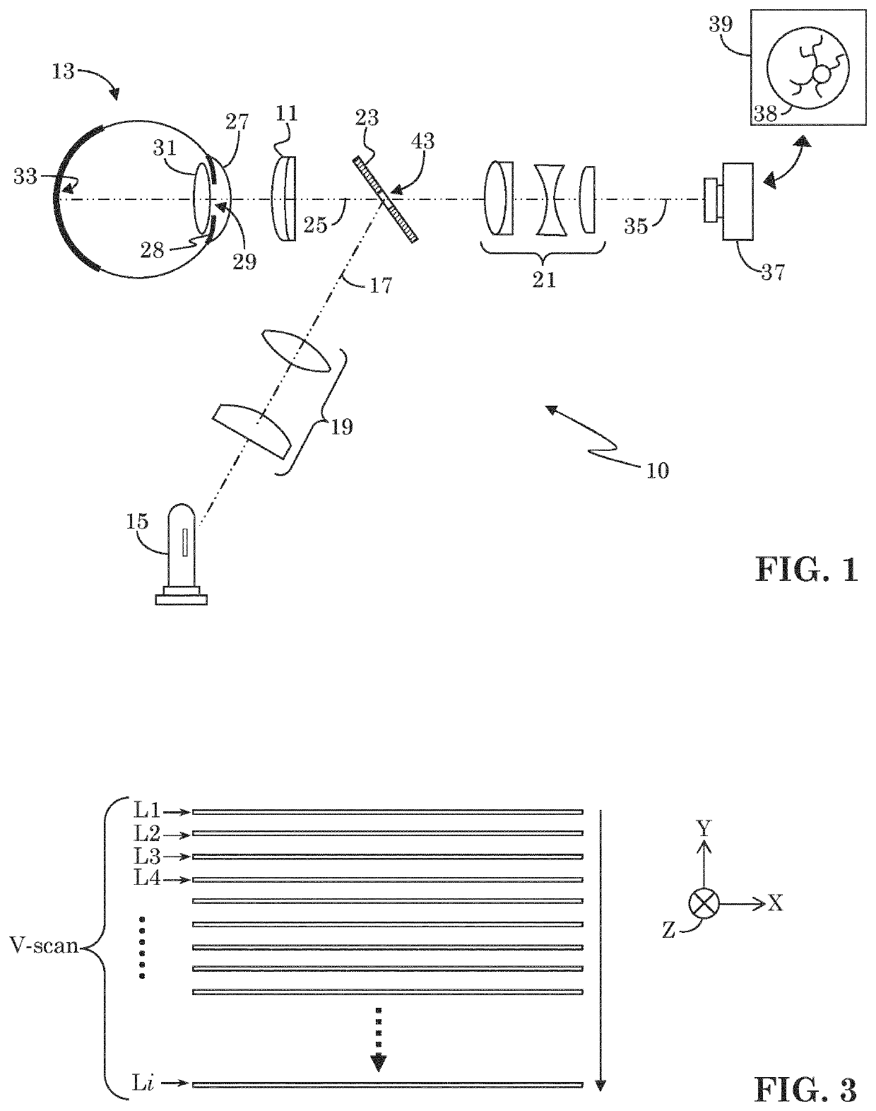
Figure 1 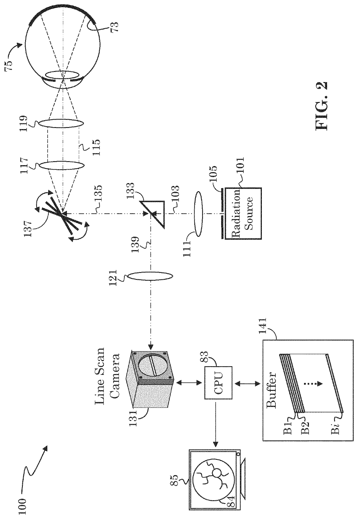
Figure 2 
Figure 3
Abstract
Description
FIELD OF INVENTION
[0001] The present invention is generally directed to the field of fundus imagers. More specifically, it is directed to a low cost fundus imager of improved field of view.BACKGROUND
[0002] Various different types of image-capture devices for imaging a sample under test are known. Of particular interest are imaging systems capable of taking close-up images of a specimen with sufficient detail, e.g., sufficient focus, lighting, magnification, and signal-to-noise ratio (SNR). An example of such an imaging system is a fundus imager, which is typically used to image the fundus of an eye. The fundus is the interior surface of the eye opposite the eye lens (or crystalline lens) and may include the retina, optic disc, macula, fovea, and posterior pole. Two categories of imaging systems used to image the fundus are flood illumination imagers and scan imagers. Scan imagers may further be divided into confocal point scanning fundus imagers and line scanning imagers.
[0003] Flood i...
Examples
Embodiment Construction
[0047]Fundus imaging is a well-established diagnostic tool in ophthalmology. A fundus camera delivers the illumination light to the back of the eye and collects the light that is reflected back. The primary challenge that any fundus camera has to overcome is to create an artifact-free image by avoiding or eliminating reflections off lenses in the fundus camera and the cornea of the human eye.
[0048]A secondary task of the fundus camera system is to aid in patient alignment or pupil alignment when coupled with another ophthalmic instrument. For example, an optical coherence tomography (OCT) system may include a fundus camera, and may make use of the fundus camera to assure proper alignment of the patient and the OCT system prior to initiating an OCT scanning operation. That is, the fundus camera may provide a real time view of the patient's eye which an OCT operator may use to assure that a desired area of the fundus is in view prior to activating an OCT scanning operation.
[0049]As st...