
Anti-block catheter
Patent Information
- Authority / Receiving Office
- US · United States
- Current Assignee / Owner
- Publication Date
- 2006-05-02
- Estimated Expiration
- Not applicable · inactive patent
Smart Images
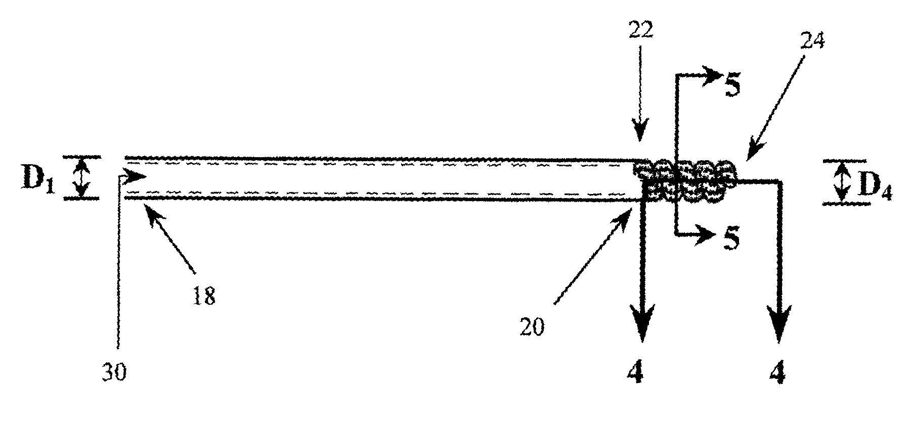
Figure 1 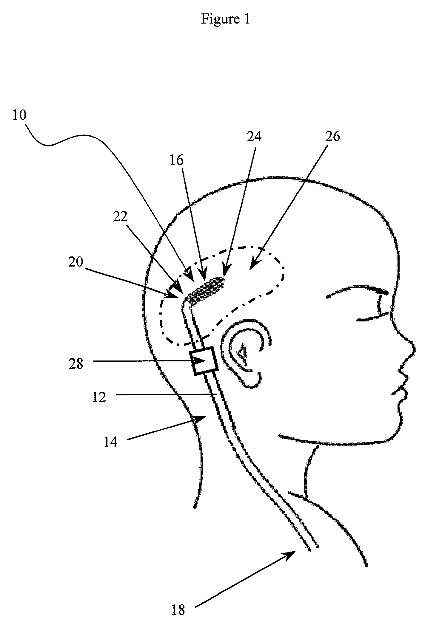
Figure 2 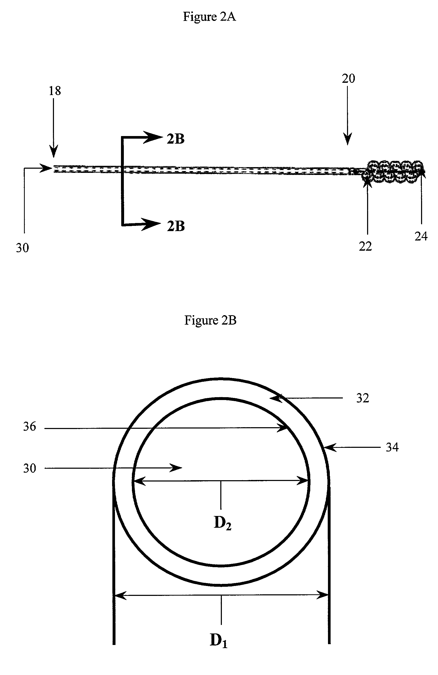
Figure 3
Abstract
Description
FIELD OF THE INVENTION
[0001] The present invention relates generally to a fluid management device that minimizes the risk of blockage or obstruction of the catheter pores, and more particularly, to a fluid management device with a catheter that includes a coiled portion having pores on the internal surface of the coil.BACKGROUND OF THE INVENTION
[0002] Hydrocephalus is a neurological condition that is caused by the abnormal accumulation of cerebrospinal fluid (CSF) within the ventricles, or cavities, of the brain. CSF is a clear, colorless fluid that is primarily produced by the choroid plexus and surrounds the brain and spinal cord. CSF constantly circulates through the ventricular system of the brain and is ultimately absorbed into the bloodstream. CSF aids in the protection of the brain and spinal cord. Because CSF keeps the brain and spinal cord buoyant, it acts as a protective cushion or “shock absorber” to prevent injuries to the central nervous system.
[0003] Hydrocephalus, which ...
Examples
Embodiment Construction
[0020]The implantable fluid management device of the present invention facilitates the removal or introduction of fluid to a treatment site. By way of example, a distal portion of the device can be implanted in the ventricle of the brain to drain excess fluid from within the ventricles, directing it to another site in the patient's body. The fluid management device of the present invention is advantageous as its construction enables fluid to be drained while minimizing the risk that the drainage catheter will be blocked or occluded.
[0021]As shown in FIG. 1, the implantable fluid management device 10 includes a catheter 12 having proximal and distal portions 14, 16 with a fluid passageway extending therebetween. The proximal portion 14 of the catheter 12 has a first, open end 18 and a transition end 20 and a lumen 30 (FIGS. 2A, 3A and 3B) connecting the two ends. The distal portion 16 of the catheter 12 is defined by an outer wall 44 (FIGS. 4 and 5) and has a first, connecting end 22...