A kind of polypeptide used for in vivo and in vitro apoptosis imaging probe and its application

An imaging probe and imaging technology, applied in the field of biomedicine, can solve the problems of radioactive pollution, high synthesis cost, unfavorable scientific research and clinical promotion, etc., and achieve the effect of easy removal and significant binding ability

Active Publication Date: 2019-01-25
THE NAT CENT FOR NANOSCI & TECH NCNST OF CHINA +1
View PDF3 Cites 0 Cited by
  • Summary
  • Abstract
  • Description
  • Claims
  • Application Information

AI Technical Summary

Problems solved by technology

At the same time, radionuclide labeling is used for imaging, not only the synthesis cost is relatively high, but also due to radioactive pollution, there are strict restrictions on the experimental environment, which is not conducive to scientific research and clinical promotion

Method used


Image

  • A kind of polypeptide used for in vivo and in vitro apoptosis imaging probe and its application
  • A kind of polypeptide used for in vivo and in vitro apoptosis imaging probe and its application
  • A kind of polypeptide used for in vivo and in vitro apoptosis imaging probe and its application

Examples


Embodiment 1

[0048] Embodiment 1: the synthesis of P17 polypeptide

[0049] The P17 polypeptide (synthesized by Shanghai Keyept Biotechnology Co., Ltd., with a purity of 98%) was synthesized according to the following sequence, and a mother solution with a suitable concentration was prepared before the experiment.

[0050] GDQNLQGPMLQGDPGFQRCIDGNVRLVFLFRG

Embodiment 2

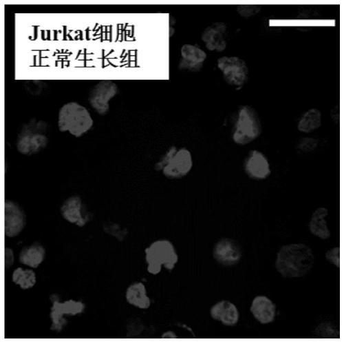
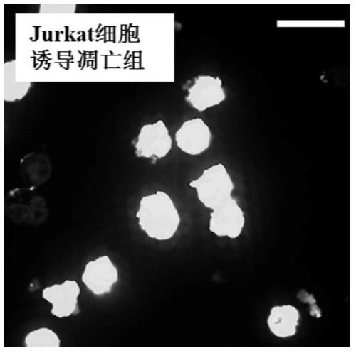
[0051] Example 2: Apoptosis imaging of P17 polypeptide probe on Jurkat cell model

[0052] Add 10 ng / mL TRAIL (TNF-related apoptosis-inducing ligand) protein to the normally growing Jurkat cell culture system, and incubate at 37°C for 24 hours to induce cell apoptosis. Add 10M fluorescein isothiocyanate (FITC)-labeled P17 polypeptide (FITC-P17) to the normal growth group and the induced apoptosis group respectively, and incubate at 37°C for 2h. Cells were washed with phosphate buffered saline (PBS) to remove unbound P17 polypeptide, and nuclei were stained with the nuclear dye 4',6-diamidino-2-phenylindole (DAPI).

[0053] Observation with laser confocal microscope, such as Figures 1A-1B As shown, under the excitation of 488nm, the green fluorescence of P17 can be seen; under the excitation of 405nm, the blue fluorescence of the cell nucleus can be seen. It shows that the P17 polypeptide has a significant binding ability to the cells after induction of apoptosis, but very li...

Embodiment 3

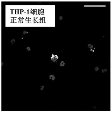
[0054] Example 3: Apoptosis imaging of P17 polypeptide probe on THP-1 cell model

[0055] Add 100ng / mL TRAIL protein to the normal growing THP-1 cell culture system and incubate at 37°C for 24h to induce cell apoptosis. Add 10M fluorescein isothiocyanate (FITC)-labeled P17 polypeptide (FITC-P17) to the normal growth group and the induced apoptosis group respectively, and incubate at 37°C for 2h. Cells were washed with PBS to remove unbound P17, after which nuclei were stained with DAPI.

[0056] Observation with laser confocal microscope, such as Figure 1C-1D As shown, under the excitation of 488nm, the green fluorescence of P17 can be seen; under the excitation of 405nm, the blue fluorescence of the cell nucleus can be seen. It shows that the P17 polypeptide has a significant binding ability to the cells after induction of apoptosis, but very little binding to the cells of normal growth.


PUM


Abstract

The invention relates to a polypeptide for in vitro and in vivo apoptosis imaging probe and an application thereof. The polypeptide has an amino acid sequence shown as SEQ ID NO: 1; and the polypeptide has significant binding capacity for induced apoptosis cells, and can specifically bind on HSP 60 protein that massively gathers in apoptotic cell cytoplasm. The polypeptide provided by the invention can be applied to in vitro apoptosis imaging or in vivo apoptosis staining; and the polypeptide probe is easy to remove and provides important new technology and new method for research and treatment for diseases such as tumors.

Description

technical field [0001] The invention relates to the technical field of biomedicine, in particular to a polypeptide and its application, in particular to a polypeptide used as an apoptosis imaging probe in vivo and in vitro and its application. Background technique [0002] Apoptosis is the most common way of programmed cell death. It not only plays an important role in physiological processes such as growth and development, but is also closely related to the occurrence, development and treatment of many diseases. For example, excessive apoptosis of cardiomyocytes in the heart is an important cause of cardiovascular diseases such as heart failure; and too little apoptosis in tissues may also lead to the occurrence of proliferative diseases, cancer, etc. What is more noteworthy is that current studies have shown that during cancer treatment, chemotherapy and radiotherapy can cause a significant increase in apoptosis in tumor tissue, and this treatment-induced apoptosis can be ...

Claims


Application Information

Patent Timeline
Patent Type & Authority Patents(China)
IPC IPC(8): C07K14/00C12N15/11C12N15/63G01N33/68A61K38/16A61P35/00A61K49/00A61P35/02
CPCA61K38/00A61K49/0006C07K14/00G01N33/68
Inventor 王琛许海燕孟洁杨棽刘健段鸿洋杨延莲
Owner THE NAT CENT FOR NANOSCI & TECH NCNST OF CHINA
Why Eureka
Try Eureka