Process for isolating vascular endothelial cells from embryoid bodies differentiated from embryonc stem cells

a technology of vascular endothelial cells and embryoid bodies, which is applied in the field of isolating vascular endothelial cells from embryoid bodies differentiated from embryonic stem cells, can solve the problems of large amount of cells being destroyed, unsatisfactory methods capable of selectively and efficiently isolating only target cells, and no effective treatment has yet been identified. , to achieve the effect of selective and high yield

Active Publication Date: 2011-04-05
CHA BIOTECH +1
View PDF5 Cites 0 Cited by
  • Summary
  • Abstract
  • Description
  • Claims
  • Application Information

AI Technical Summary

Benefits of technology

[0011]While searching for a high yield and selective method for isolating vascular endothelial cells differentiated from human embryonic stem cells, the present inventors have surprisingly found that when embryoid bodies differentiated from human embryonic stem cells are subjected to a two-step treatment of trypsin and ethylenediaminetetraacetate (EDTA) including a lower dose trypsin-EDTA treatment and a higher dose trypsin-EDTA treatment, specific regions of the embryoid bodies containing only a trace amount of vascular endothelial cells can be removed easily and rapidly, and the isolated vascular endothelial cells retain their intrinsic characteristics.

Problems solved by technology

Cardiovascular diseases are considered to be very serious diseases that cost the United States US$ 260 billion every year, but no effective treatment has yet been identified.
However, satisfactory methods capable of selectively and efficiently isolating only target cells have not yet been reported.
However, while isolating single cells from differentiated embryoid bodies using FACS, large amounts of the cells are destroyed, and further, the recovery rate of vascular endothelial cells is merely 2%.

Method used


Image

  • Process for isolating vascular endothelial cells from embryoid bodies differentiated from embryonc stem cells
  • Process for isolating vascular endothelial cells from embryoid bodies differentiated from embryonc stem cells
  • Process for isolating vascular endothelial cells from embryoid bodies differentiated from embryonc stem cells

Examples


example 1

[0034]Mouse fibroblasts (STO cells, 2.5×10⁵ cells / well), which had been treated with mitomycin-C for two hours to prevent cell proliferation, and feeder cell culture media (90% DMEM supplemented with 10% FBS, 0.1 mM mercaptoethanol, and 1% nonessential amino acid (Gibco)) were plated on gelatin-coated culture dishes, and the STO cells were cultured for 24 hours. The culture dishes were washed twice with phosphate is buffered saline media. The media in the culture dishes were replaced with culture media (80% KO-DMEM supplemented with 20% serum replacement (SR), 0.1 mM mercaptoethanol, 1% nonessential amino acid (Gibco), and 4 ng / ml bFGF), and human embryonic stem cells (CHA-hES3) were cultured for about seven days.

[0035]Human embryonic stem cell colonies formed in the culture media were detached from neighboring feeder cells by a glass pipette, and were then suspension-cultured in culture media including 20% serum replacement (Gibco), 0.1 mM mercaptoethanol, 1% nonessential amino aci...

example 2

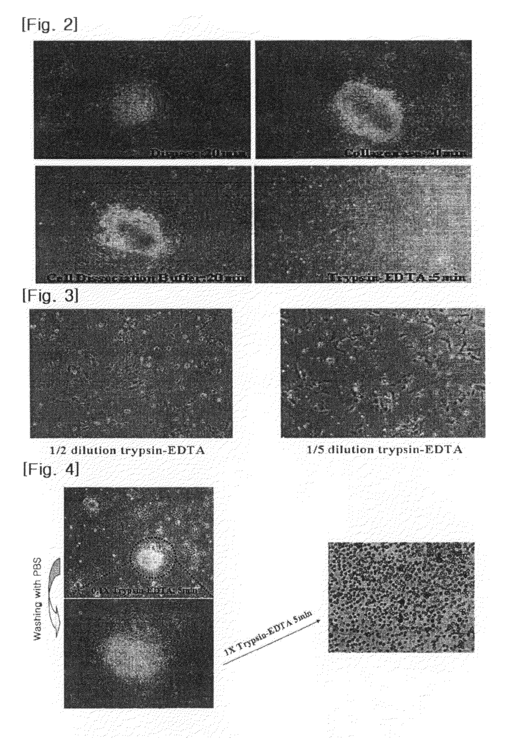
Step 1: Lower Dose Trypsin-EDTA Treatment

[0037]The embryoid bodies obtained in Example 1 were seeded on culture media (90% DMEM supplemented with 10% FBS, 0.1 mM mercaptoethanol, and 1% nonessential amino acid (Gibco)). A 10-fold dilute solution of a trypsin-EDTA solution (Sigma, U.S.A.) of 0.25% trypsin and 1 mM EDTA in a 0.9% sodium chloride solution was added to the culture media, and the embryoid bodies were cultured for five minutes.

Step 2: Separation into Single Cells

[0038]The embryoid bodies obtained in step 1 were washed with a phosphate buffered saline to thereby completely remove cell clusters in the outer regions of the suspended embryoid bodies. A trypsin-EDTA solution (Sigma, U.S.A.) of 0.25% trypsin and 1 mM EDTA in a 0.9% sodium chloride solution was added, and the remaining cell clusters were cultured for five minutes to thereby separate the cell clusters into single cells.

example 3

[0039]The same treatment as in Example 2 was performed except that in step 1, a 8-fold dilute solution of the trypsin-EDTA solution (Sigma, U.S.A.) was added to embryoid bodies, and the embryoid bodies were cultured for one, three, and five minutes.


PUM

PropertyMeasurementUnit
concentrationaaaaaaaaaa
cell massaaaaaaaaaa
Fluorescence Activated Cell Sorteraaaaaaaaaa

Abstract

The present invention provides a process for isolating vascular endothelial cells from embryoid bodies differentiated from embryonic stem cells, which comprises: (a) treating embryoid bodies differentiated from embryonic stem cells with 0.005-0.015% trypsin and 0.05-0.15 mM ethylenediaminetetraacetate (EDTA) to obtain vascular endothelial cell clusters; and (b) treating the vascular endothelial cell clusters with 0.1-0.5% trypsin and 0.5-2 mM EDTA so as to separate the vascular endothelial cell clusters into single cells.

Description

CROSS REFERENCE TO RELATED APPLICATIONS[0001]This application is a 35 USC §371 National Phase Entry Application from PCT / KR2007 / 002840, filed Jun. 13, 2007, and designating the United States.TECHNICAL FIELD[0002]The present invention relates to a process for isolating vascular endothelial cells from embryoid bodies differentiated from embryonic stem cells.BACKGROUND ART[0003]Cardiovascular diseases are one of the leading causes of death worldwide. It is estimated that every year, about 12 million people throughout the world die due to cardiovascular diseases. Cardiovascular diseases are considered to be very serious diseases that cost the United States US$ 260 billion every year, but no effective treatment has yet been identified.[0004]Recently, studies have been reported on inducing the differentiation of human embryonic stem cells into vascular endothelial cells, muscle cells, etc., raising hope for the possibility of treatment of cardiovascular diseases using human embryonic stem...

Claims


Application Information

Patent Timeline
Patent Type & Authority Patents(United States)
IPC IPC(8): C12N5/02C12N5/071
CPCC12N5/069C12N2509/00C12N2506/02
Inventor CHUNG, HYUNG-MINMOON, SUNG-HWANKIM, JU-MILEE, SOO-HONG
Owner CHA BIOTECH
Why Eureka
Try Eureka