Device for performing examination through the uterine cavity

a technology for uterine cavity and uterine cavity, which is applied in the field of medical devices, can solve the problems of increasing the trauma experience of the patient, deficient procedures, and considerable time-consuming, and achieves the effect of convenient and rapid assembly and operation

Active Publication Date: 2016-06-07
CARRASCOSA PATRICIA MARINA
View PDF6 Cites 0 Cited by
  • Summary
  • Abstract
  • Description
  • Claims
  • Application Information

AI Technical Summary

Benefits of technology

[0011]It is therefore a purpose of the current invention to provide a new, simple device that can easily and quickly be assembled and operated for the preparation of a patient for medical examination of the uterine cavity which does not require traction of the cervix as it can be applied with digital imagery technology such as multi-slice tomographs and other modern imagery systems.

Problems solved by technology

This item is particularly important for the trauma that is caused to the patient, using a forceps that grabs the cervix, not only because it causes discomfort, in the best of cases, but also because it can be painful and causes small wounds with corresponding bleeding and / or additional infections.
This procedure is deficient because of at least three significant disadvantages.
One is that it is considerably time-consuming, at times up to 40 minutes, which only increase the traumatic experience for the patient.
Another disadvantage is that the cannula, grasper, connections of the cannula with the source of the liquid and the manual handles for the injection of the liquid into the uterus should be held and manipulated by professional staff that must stay with the patient almost constantly, whereby they are almost always exposed to X-radiation.
Finally, the other disadvantage is that it only provides information about the interior of the organs (cervix, uterus and tubes), a “luminogram”, without giving information about the wall or their external morphology.

Method used


Image

  • Device for performing examination through the uterine cavity
  • Device for performing examination through the uterine cavity
  • Device for performing examination through the uterine cavity

Examples


Embodiment Construction

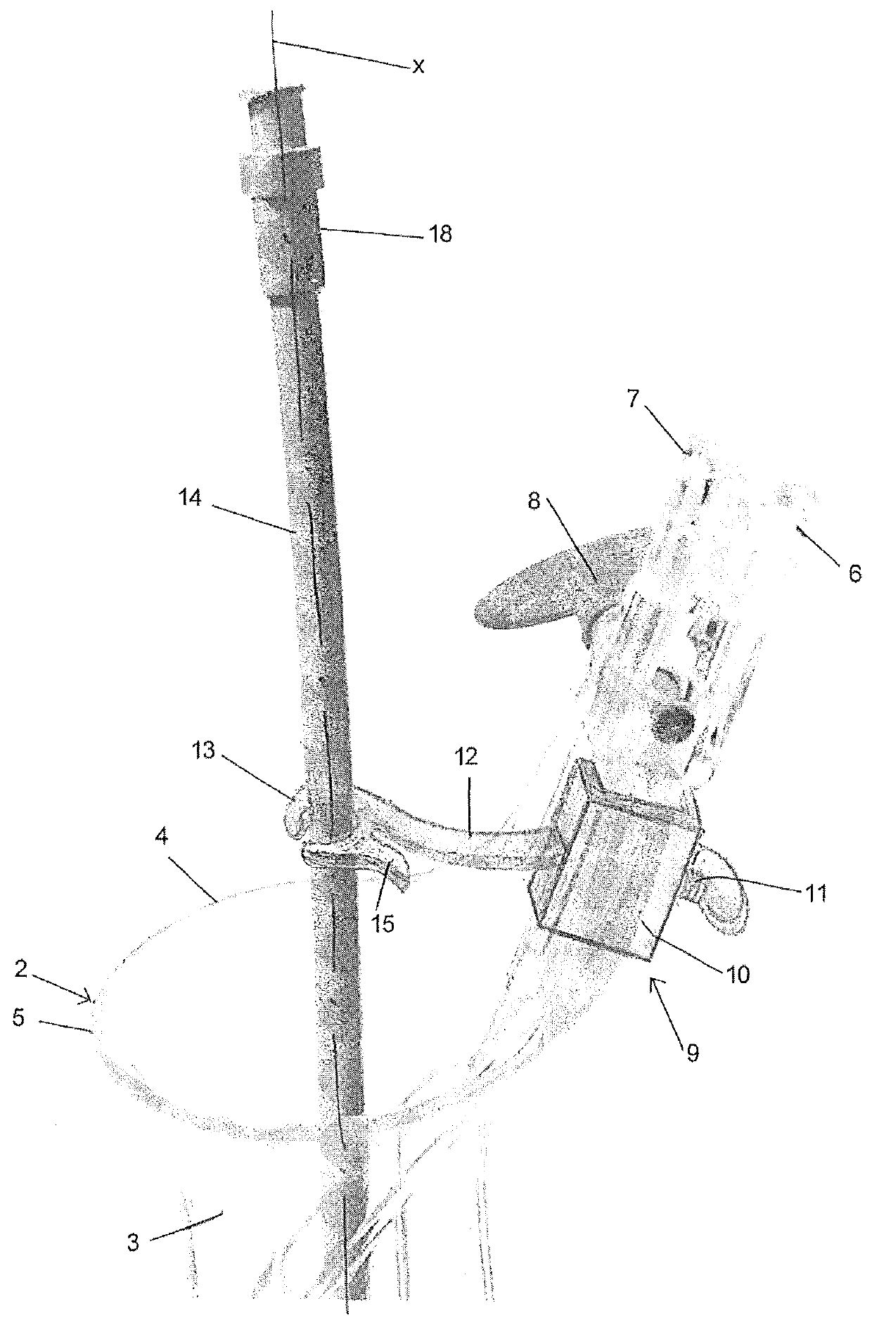
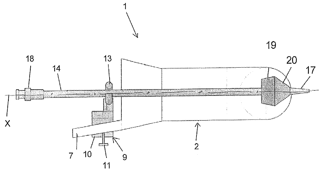
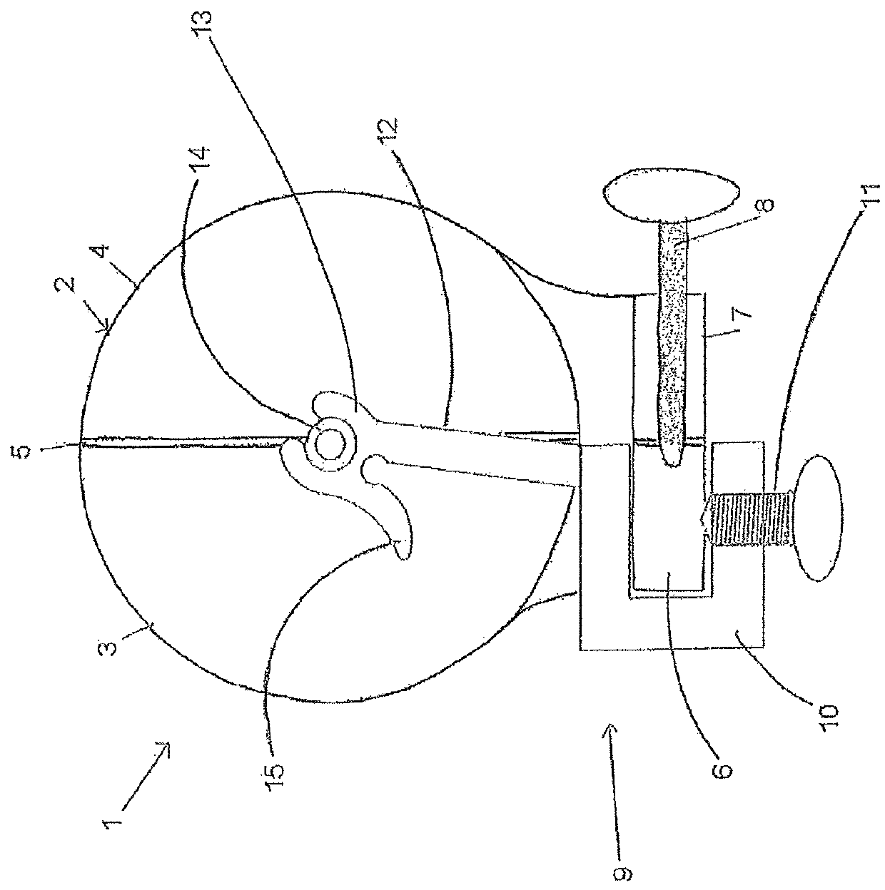
[0019]By looking at the figures we can deduce that the invention comprises a device for performing an examination through the uterine cavity, and more preferably to perform a procedure corresponding to an examination known as computed tomography virtual hysterosalpingography, which has been referenced above in this description. The device, indicated with the general reference number 1 in the drawings, is composed by a speculum (2) that can comprise two or more valves and that preferably is a speculum composed by two valves 3 and 4, opposed by their sides, as indicated with reference number 5. The speculum 2 can be a known speculum, whose valves 3 and 4 are joined together by an opening / closing mechanism with an adjustable hinge. The valve opening / closing mechanism comprises two arms 6, 7 that are connected by means of an internal hinge (which has not been illustrated) and an adjustment screw 8 which is screwed onto at least one of said arms of the speculum. By handling the screw 8 i...


PUM


Abstract

A device for performing examination through the uterine cavity, preferably to carry out a computed tomography virtual hysterosalpingography, by the use of a speculum and a cannula or probe, wherein the device comprises a support for centering in the speculum and the probe is retained in a central end of the support to guarantee the centering of the probe within the cervix and the holding of the probe in the desired position during the test.

Description

CROSS-REFERENCE TO RELATED APPLICATIONS[0001]This Application is a U.S. National Stage application under 35 U.S.C. 371 of International Application No. PCT / ES2010 / 070214 which claims priority to Argentine Application AR090104134, filed Oct. 27, 2009, each of which is hereby incorporated by reference herein in its entirety.BACKGROUND OF THE INVENTION[0002]1. Field of the Invention[0003]The current invention is related to the field of medical devices used for medical exams related to the feminine genital organ and, more precisely, reference is made to a device to perform an examination called Multidetector computed tomography virtual hysterosalpingography that allows for the use of digital technology to take images and avoid traumatic manipulations to which the patient is submitted with radiologic technologies and associated devices that are currently being used.[0004]2. Description of the Prior Art[0005]Before starting off with the description of the devices and systems that are curr...

Claims


Application Information

Patent Timeline
Patent Type & Authority Patents(United States)
IPC IPC(8): A61B1/32A61B1/303A61B17/02A61B17/42
CPCA61B1/303A61B17/0206A61B17/42
Inventor CARRASCOSA, PATRICIA MARINA
Owner CARRASCOSA PATRICIA MARINA
Why Eureka
Try Eureka