
Immunoreactive francisella tularensis antigens
Patent Information
- Authority / Receiving Office
- US · United States
- Current Assignee / Owner
- Publication Date
- 2013-07-18
- Estimated Expiration
- Not applicable · inactive patent
Smart Images
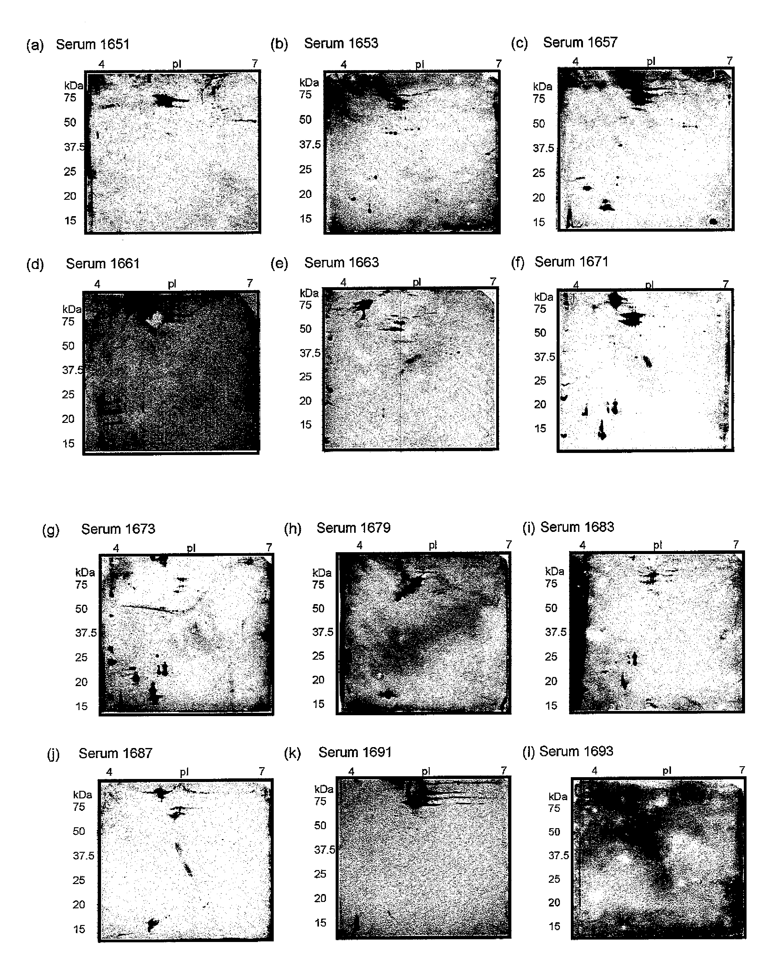
Figure 1 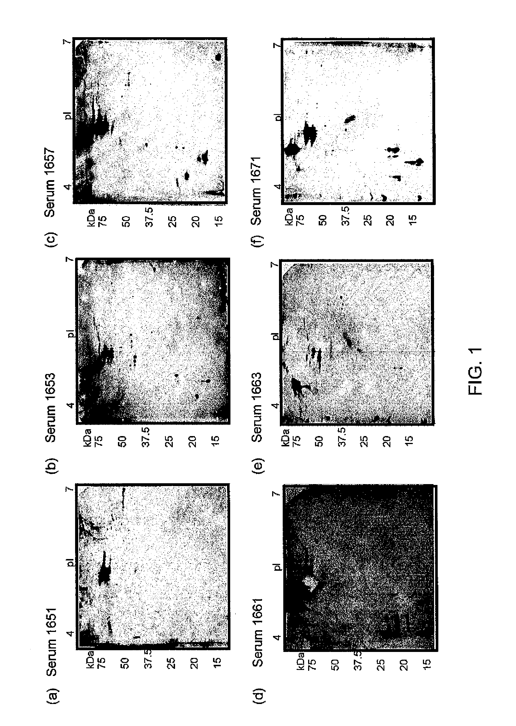
Figure 2 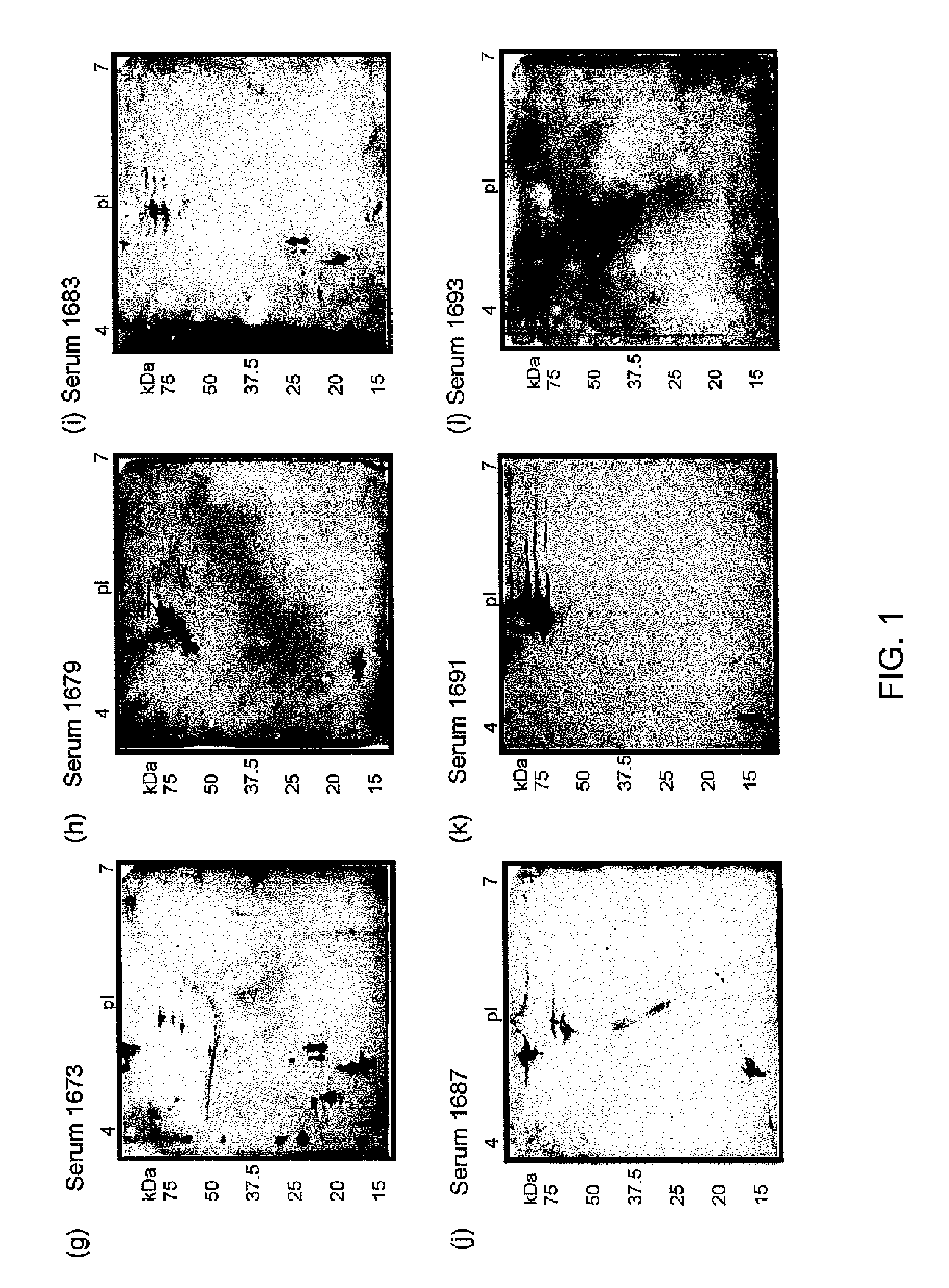
Figure 3
Abstract
Description
FIELD OF THE INVENTION
[0001] The present invention relates to immunoreactive Francisella tularensis antigens and uses thereof. More specifically, the invention relates to immunoreactive Francisella tularensis antigens and their uses as correlates of protection.BACKGROUND OF THE INVENTION
[0002] Tularemia is a disease caused by the Gram-negative facultative intracellular bacterium, Francisella tularensis. F. tularensis is pathogenic for many mammalian species including humans, causing a spectrum of diseases collectively called tularemia. Tularemia has been reported as a clinical infection in primates in many temperate climates across the world. Several subspecies exist, with the most clinically relevant subspecies being holarctica and tularensis, commonly denoted Type B and A strains, respectively (Sjostedt, 2001). The subspecies tularensis (Type A) is endemic only to North America. Mortality rates of up to 60% have been reported for untreated human cases of disseminated infection cause...
Examples
example 1
Sera Preparations
[0066]Four distinct collections of human sera were used in Examples 2 and 3, including human tularemia patients (two separate groups), LVS-vaccinated laboratory personnel, and clinical trial subjects immunized with LVS. The details of each sera screened are shown in Table 2 and summarized briefly below.
[0067]The Type B convalescent sera were obtained from patients diagnosed with tularemia in a region of Sweden, where the disease is considered endemic. Control sera were obtained from individuals with no history of tularemia or a tularemia-like disease. In total, sera were obtained from 12 tularemia patients and 3 healthy individuals with no history of tularemia. Since Type A strains are endemic to North America only, the Swedish patients were exclusively infected with type B strains. The route of infection for the majority of these patients was intradermal.
[0068]The Type A convalescent sera were obtained from a subset of 59 subjects with presumed or confirmed cases o...
example 2
Two-Dimensional Polyacrylamide Gel Electrophoresis Western Blotting
[0071]The four collections of human sera described in Example 1 were used in two-dimensional polyacrylamide gel electrophoresis (2D PAGE) Western blotting experiments in order to determine the repertoire of immunoreactive proteins for each serum sample. Briefly, the proteins of a bacterial cell lysate were separated in two dimensions—by protein isoelectric point then by protein molecular mass using 2D-PAGE, as described in Twine et al (2005; 2010). Resolved proteins were then transferred to nitrocellullose membrane by electroblotting and the membrane was subsequently incubated with serum from Example 1. Antibodies in the serum recognised their cognate antigen on the membrane and this antibody binding was subsequently detected, generating a pattern of immunoreactive proteins.
[0072]Francisella tularensis Δwbtl, a mutant strain lacking the O-antigen, was used as the protein antigen in blotting experiments. Briefly, bact...
example 3
Identification of Immunoreactive Proteins
[0076]Immunoreactive proteins observed by 2D Western blotting in Example 2 were identified by tryptic digest and mass spectrometry.
[0077]Protein spots corresponding to areas of immunoreactivity on Western blots were excised from equivalent protein stained 2D-PAGE gels and tryptically digested manually, as described previously (Twine et al, 2010; Twine et al, 2006). The in-gel digests were analyzed by nano-liquid chromatography-MS / MS as described previously (Twine et al, 2010). The peak list files of MS2 spectra of the excised protein spots were searched against the translated SCHU S4 genome sequence using the MASCOT™ search engine (version 2.2.03) (Matrix Science, London, UK) for protein identification, as described in earlier work (Twine et al, 2010).
[0078]A total of 31 immunoreactive proteins were identified from Type B patients (Table 3, FIG. 6). A single protein, Chaperonin GroEL (FTT—1696), was immunoreactive with all sera from all 12 pa...