
Endoscope Tip Part and Endoscope
Patent Information
- Authority / Receiving Office
- US · United States
- Current Assignee / Owner
- Publication Date
- 2014-03-20
Smart Images
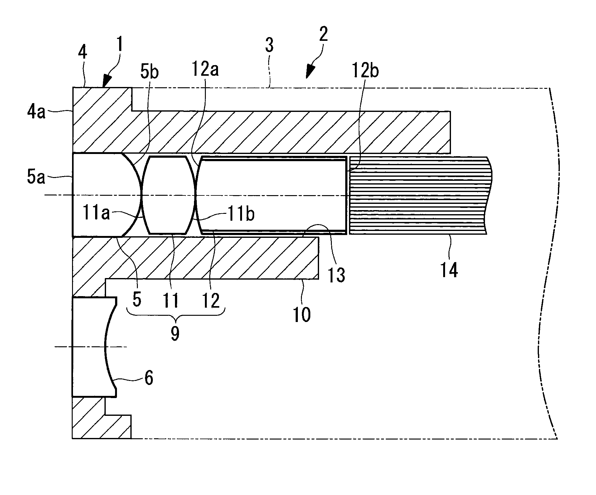
Figure 1 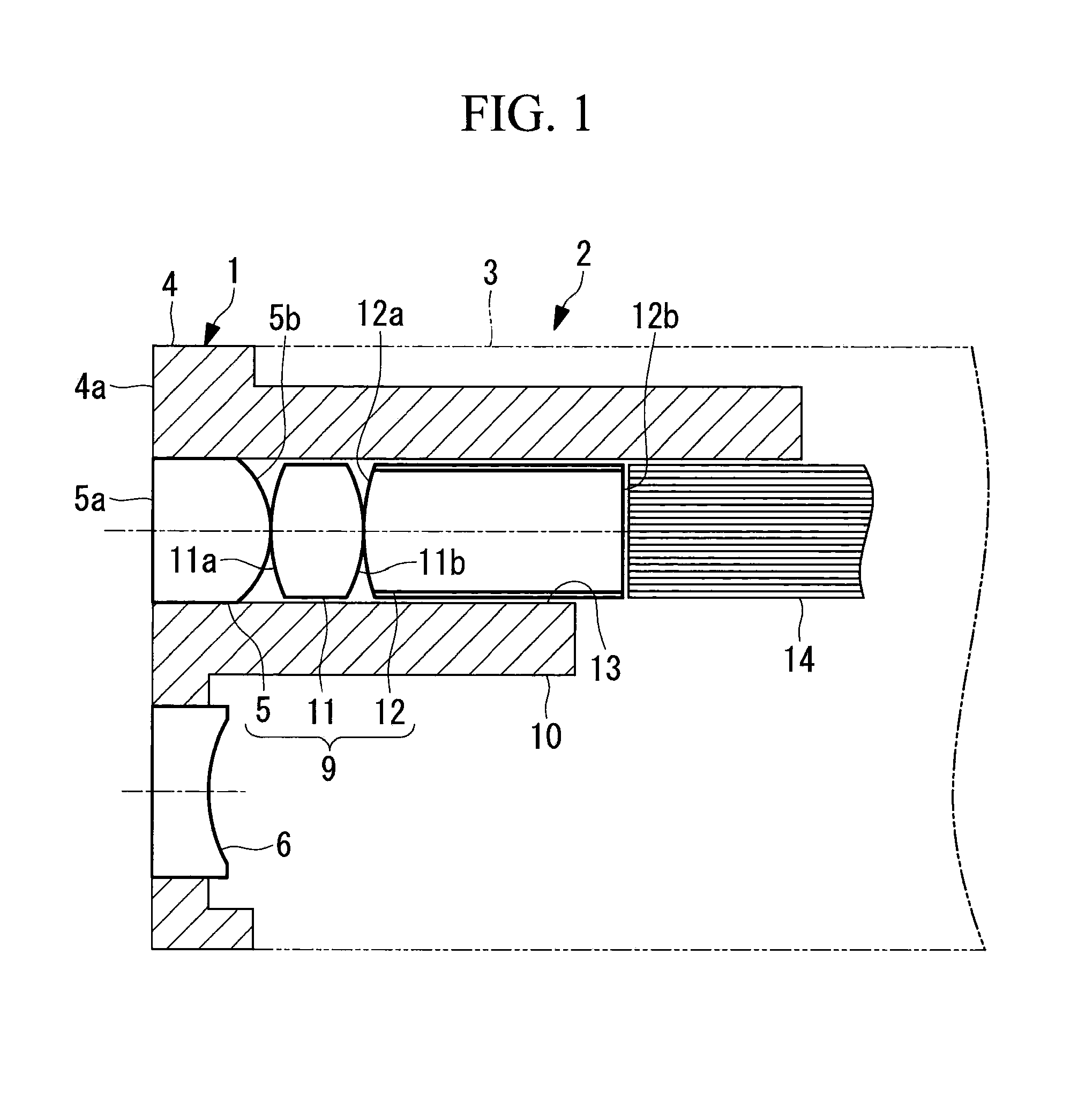
Figure 2 
Figure 3
Abstract
Description
CROSS-REFERENCE TO RELATED APPLICATIONS
[0001] This is a continuation of International Application PCT / JP2012 / 081886, with an international filing date of Dec. 10, 2012, which is hereby incorporated by reference herein in its entirety. This application is based on Japanese Patent Application No. 2012-005293, the contents of which are incorporated herein by reference.TECHNICAL FIELD
[0002] The present invention relates to an endoscope tip part and an endoscope.BACKGROUND ART
[0003] In general, an objective optical system for acquiring images of an observation object and an illumination optical system for irradiating the observation object are arranged at the tip portion of an insertion section of an endoscope. Furthermore, an imaging device that acquires images of the observation object imaged by the objective optical system is installed in the insertion section.
[0004] Furthermore, a first lens of the objective optical system and a first lens of the illumination optical system are disposed ...
Examples
example 1
[0084]FIG. 5 shows the configuration of an illumination optical system 9 of the endoscope 2 according to this example, and Table 1 shows values of design data. The illumination optical system 9 is formed of, in order from the object side, the first lens 5 having positive refractive power, the second lens 11 having positive refractive power, and the third lens 12 having positive refractive index.
[0085]Note that the third lens 12 is a rod lens, and the surface thereof closer to the light source is an end surface to which light is emitted from the light guide fibers 14. The first lens 5 is integrated with the tip part main body 4 through two-color molding. Furthermore, the rear second lens 11 and third lens 12 are assembled by being butted against each other at the centers of the optical axes of the lenses.
[0086]Thus, the distance between the lens surfaces is 0. With this lens assembly method, spacing members for the lenses 5, 11, and 12 are not required, which achieves a reduction in ...
example 2
[0088]Table 2 shows values of design data of an illumination optical system 9 of the endoscope 2 according to this example. This illumination optical system 9 has the same configuration as that of Example 1 and is equivalent in terms of the assembly method, performance, and advantageous effects.
TABLE 2radius ofAbbelenssurfacecurvatureintersurfacerefractivenumberouternumberrdistance dindex Neνddiameter1∞1.651.6414723.2φ2.22−1.4503 2.381.541.8881540.76φ2.24−2.38053.68(rod)3.881.6522225.42φ2.26∞0refractive index of rod cladding: 1.514focal length: 1.00 mm
example 3
[0089]Table 3 shows values of design data of an illumination optical system 9 of the endoscope 2 according to this example. This illumination optical system 9 has the same configuration as that of Example 1 or Example 2 and is equivalent in terms of the assembly method, performance, and advantageous effects.
TABLE 3radius ofAbbelenssurfacecurvatureintersurfacerefractivenumberouternumberrdistance dindex Neνddiameter1∞1.61.6414723.2φ22−1.4303 2.781.551.8881540.76φ24−2.05053.37(rod)3.81.6522225.42φ26∞0refractive index of rod cladding: 1.514focal length: 1.00 mm
[0090]Table 4 shows values of conditional expressions (1) to (8) in the configurations of the respective examples.
TABLE 4conditionalexpressionExample 1Example 2Example 3(1)1.641.641.64(2)7.337.086.95(3)1.541.451.40(4)0.790.790.68(5)0.640.640.64(6)3.843.603.37(7)5.895.535.17(8)1.071.121.14
Additional Items
[0091]Note that, in the present invention, the following configurations can also be adopted.
Additional Item 1
[0092]An endoscope t...