Three dimensional image processing apparatus and x-ray diagnosis apparatus

Active Publication Date: 2008-06-12
TOSHIBA MEDICAL SYST CORP
View PDF6 Cites 58 Cited by
  • Summary
  • Abstract
  • Description
  • Claims
  • Application Information

AI Technical Summary

Benefits of technology

[0008]It is an object of the present invention to improve the image quality of

Problems solved by technology

Since coronary vessels move in accordance with pulsations, motion artifacts occur.
In addition, since the motion of the heart is not constant, motion correction based on a model is not sufficient, and it is necessary to perform motion correction based on an actual image.
Erroneous designation by the operator, i.e., a human error, will degrade the quality of a reconstructed image.
Many designation errors appear in a case wherein a blood vessel overlaps a region of interest (stricture or stent), a case (Foreshortening) wherein an image is displayed with an apparently reduced depth due to projection, a case wherein an object blurs due to the hard mo

Method used


Image

  • Three dimensional image processing apparatus and x-ray diagnosis apparatus
  • Three dimensional image processing apparatus and x-ray diagnosis apparatus
  • Three dimensional image processing apparatus and x-ray diagnosis apparatus

Examples


Embodiment Construction

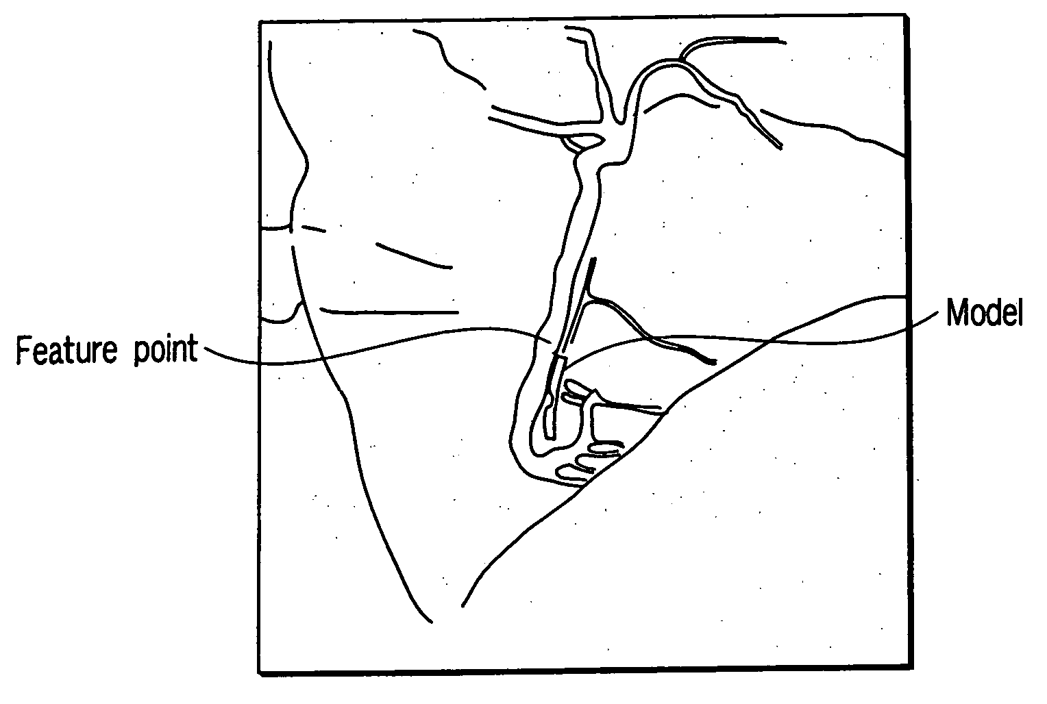
[0085]A three dimensional image processing apparatus and an X-ray diagnosis apparatus according to an embodiment of the present invention will be described below with reference to the views of the accompanying drawing. The three dimensional image processing apparatus will be described as an apparatus incorporated in a radiographic apparatus. Obviously, however, this apparatus may be singly used. Alternatively, the three dimensional image processing apparatus may be implemented as a program for causing a computer to implement the function of the apparatus, or can be provided as a computer-readable storage medium which stores the program. Although a target object will be described as a heart blood vessel, three dimensional image processing can be applied to even an organ other than a heart blood vessel or a device to be inserted in the body of a subject, e.g., a stent.

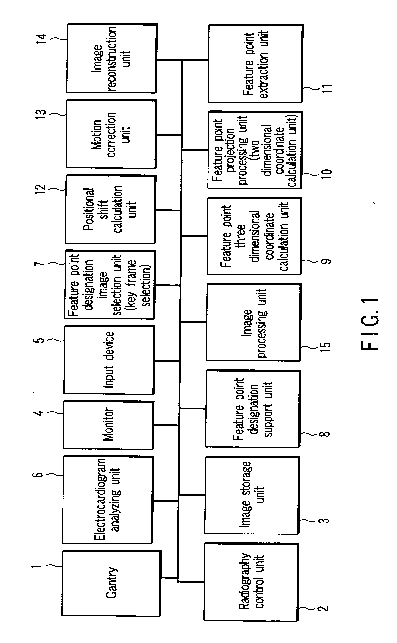
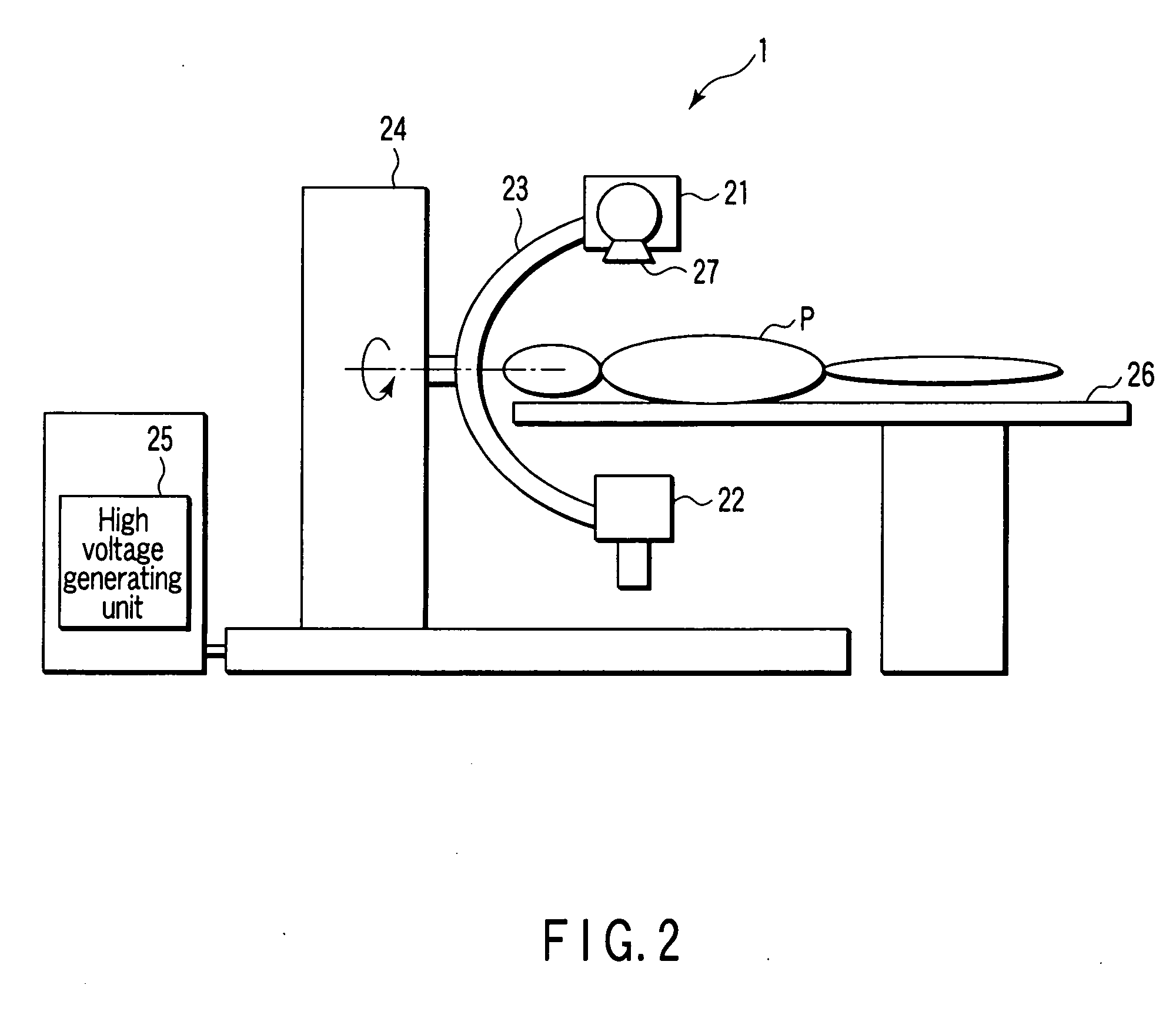
[0086]FIG. 1 shows a radiographic apparatus incorporating a three dimensional image processing apparatus according to ...


PUM


Abstract

A three dimensional image processing apparatus includes a feature point designation unit which designates feature points on at least two selected images selected from a plurality of images in different radiographing directions, a three dimensional position calculation unit which calculates a three dimensional position associated with a feature point, a two dimensional position calculation unit which calculates the two dimensional position of a feature point on an unselected image on the basis of the calculated three dimensional position of the feature point, a feature point extraction unit which extracts a feature point from an unselected image, a positional shift calculation unit which calculates a positional shift of the two dimensional position of the extracted feature point with respect to the calculated two dimensional position of the feature point, a correction unit which corrects the position of the unselected image on the basis of the calculated positional shift, and an image reconstruction unit which reconstructs a three dimensional image on the basis of the selected image and the corrected unselected image.

Description

BACKGROUND OF THE INVENTION[0001]1. Field of the Invention[0002]The present invention relates to a three dimensional image processing apparatus which reconstructs a three dimensional image from a plurality of X-ray images obtained in different radiographing directions and an X-ray diagnosis apparatus.[0003]2. Description of the Related Art[0004]Recently, studies have been made on a technique of reconstructing a three dimensional image from a plurality of X-ray images obtained in different radiographing directions using the same technique as that for X-ray computed tomography. For example, this technique is useful for cardiovascular examination. Since coronary vessels move in accordance with pulsations, motion artifacts occur.[0005]In order to solve this problem, it is necessary to correct the motions of coronary vessels. In addition, since the motion of the heart is not constant, motion correction based on a model is not sufficient, and it is necessary to perform motion correction b...

Claims


Application Information

Patent Timeline
IPC IPC(8): G06K9/00
CPCA61B6/4441A61B6/469A61B6/503G06T2211/412A61B6/5264A61B6/5288G06T11/005A61B6/504
Inventor SAKAGUCHI, TAKUYASIVER, MICHAEL D.
Owner TOSHIBA MEDICAL SYST CORP
Why Eureka
Try Eureka