
Breast cancer stem cell vaccine as well as preparation method and application thereof
A breast cancer stem cell, breast cancer cell technology, applied in the direction of tumor/cancer cells, animal cells, vertebrate cells, etc., can solve the problems of low efficacy and lack of breast cancer stem cell vaccine, etc., to enhance the efficacy, improve the body's immune function, Ease of quality control
Patent Information
- Authority / Receiving Office
- CN · China
- Patent Type
- Applications(China)
- Current Assignee / Owner
- Publication Date
- 2014-12-24
Smart Images
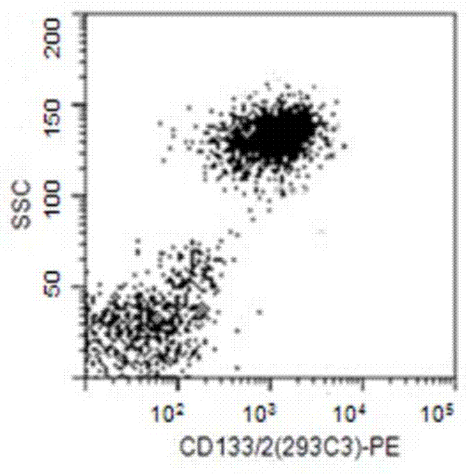
Figure 1 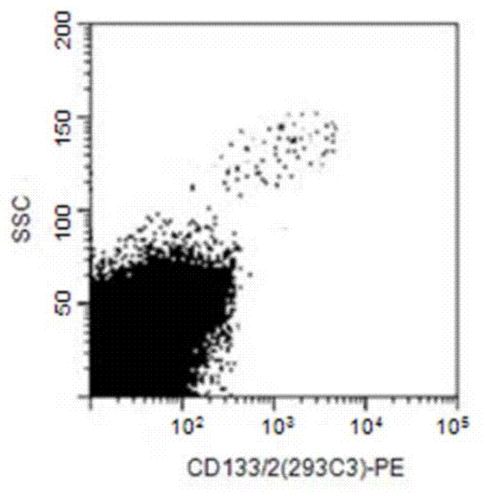
Figure 2 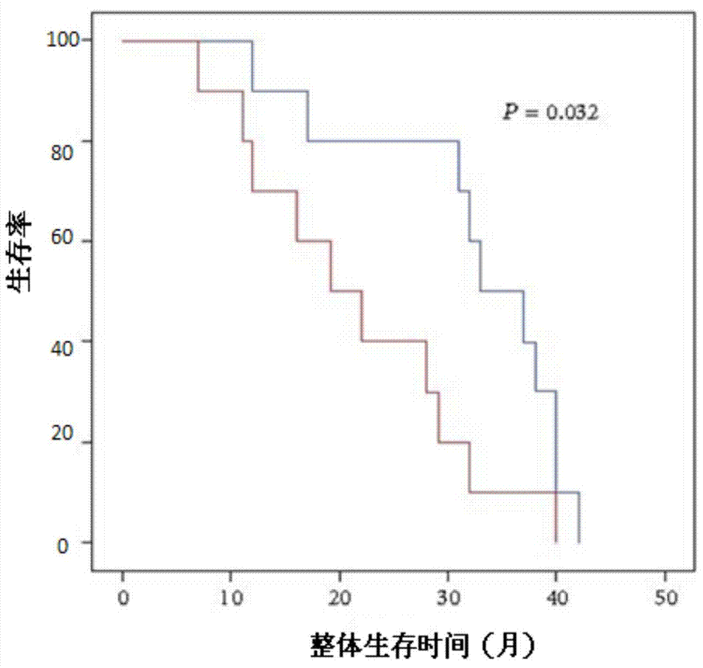
Figure 3
Abstract
Description
technical field
[0001] The invention relates to biomedical engineering technology, and relates to a tumor stem cell vaccine and its preparation and application. Background technique
[0002] Malignant tumor is a systemic chronic disease. In patients with middle and advanced stages, there are not only obvious mass tumors in the organs during imaging examinations, but also a large number of invisible circulating tumor cells and cancer stem cells (Cancer stem cells, CSCs) in the peripheral blood, ready to settle in the body at any time Susceptible sites develop into new metastases, which is why the tumor cannot be cured. Traditional tumor treatment includes 4 main therapies, namely surgery, chemotherapy, radiotherapy and immunotherapy. Usually, after 3-5 years of treatment with the first three therapies, the tumor will metastasize to peripheral organs or distant organs. With the in-depth research on CSC, it has been clear that the reason for recurrence and metastasis is not ...
Examples
Embodiment 1
[0041] Embodiment 1: Preparation of breast cancer stem cell vaccine
[0042] In this example, breast cancer stem cell vaccines were prepared using breast cancer cells stored in liquid nitrogen as raw materials.
[0043] 1. Put the breast cancer ductal cells HCC1937 stored in liquid nitrogen or -80°C in a 37°C water bath to dissolve quickly;
[0044] 2. Mix the thawed tumor cell suspension with 8.0ml medium (RPMI1640+10% FBS), and centrifuge at 1500 rpm for 8 minutes;
[0045] 3. Discard the supernatant, then absorb 8.0ml culture medium to mix the cell pellet, then centrifuge at 1500 rpm for 8 minutes, discard the supernatant, mix the cell pellet with 1.0ml culture medium, and set aside;
[0046] 4. Take another 75cm 2 Square bottle, add 14.0ml of culture medium, add the suspension containing tumor cells (1.0ml) prepared above into this square bottle, at 37°C, 5% CO 2 cultured in an incubator;
[0047] 5. Tumor cells grow on the wall. After 3-4 days, when the tumor cells gr...
Embodiment 2
[0064] Example 2: Observation of curative effect after injection of breast cancer stem cell vaccine
[0065] Apply the breast cancer stem cell vaccine prepared in Example 1 to the treatment of breast cancer patients. The detailed experimental process is as follows: 30 breast cancer patients were randomly selected and divided into two groups, 15 of which received the vaccine injection treatment prepared in Example 1. , as the treatment group; the other 15 people who did not receive vaccine treatment were used as the control group. Before injecting patients, 5ml of blood was drawn from each patient, and the number of peripheral blood lymphocytes (T BNK detection kit) and lymphocyte function (6colour T BNK With Trucount detection kit) were detected, and the report sheet was obtained through flow cytometry detection. For patients in the treatment group, inject 500ul of the vaccine into the body of the breast cancer patient subcutaneously, and inject it again after a week interval,...
Embodiment 3
[0073] Example 3: Humoral immune response induced by the tumor stem cell vaccine prepared in Example 1
[0074] 30 routine patients in the embodiment 1 are equally divided into two groups (no vaccine injection group-control group and vaccination group-experimental group), carry out according to the following method respectively:
[0075] Collect 5ml of the whole blood of the patient on the 14th day, and separate the plasma with the lymphocyte separation medium. The details are as follows: Take 5ml of fresh anticoagulant blood, mix it with normal saline for injection at a ratio of 1:1, and carefully add it to 2ml of the lymphocyte separation medium , centrifuge at 400g (horizontal rotor with a radius of 15cm) for 20 minutes. At this time, the centrifuge tube is divided into four layers, and the uppermost layer is plasma. The IgG content in plasma was detected by ELISA.
[0076] The result is as image 3 As shown, compared with the control group, P image 3 It can be seen that ...