
Scintillation detector based systems and methods for using the same
Patent Information
- Authority / Receiving Office
- US · United States
- Current Assignee / Owner
- Publication Date
- 2022-07-07
Smart Images
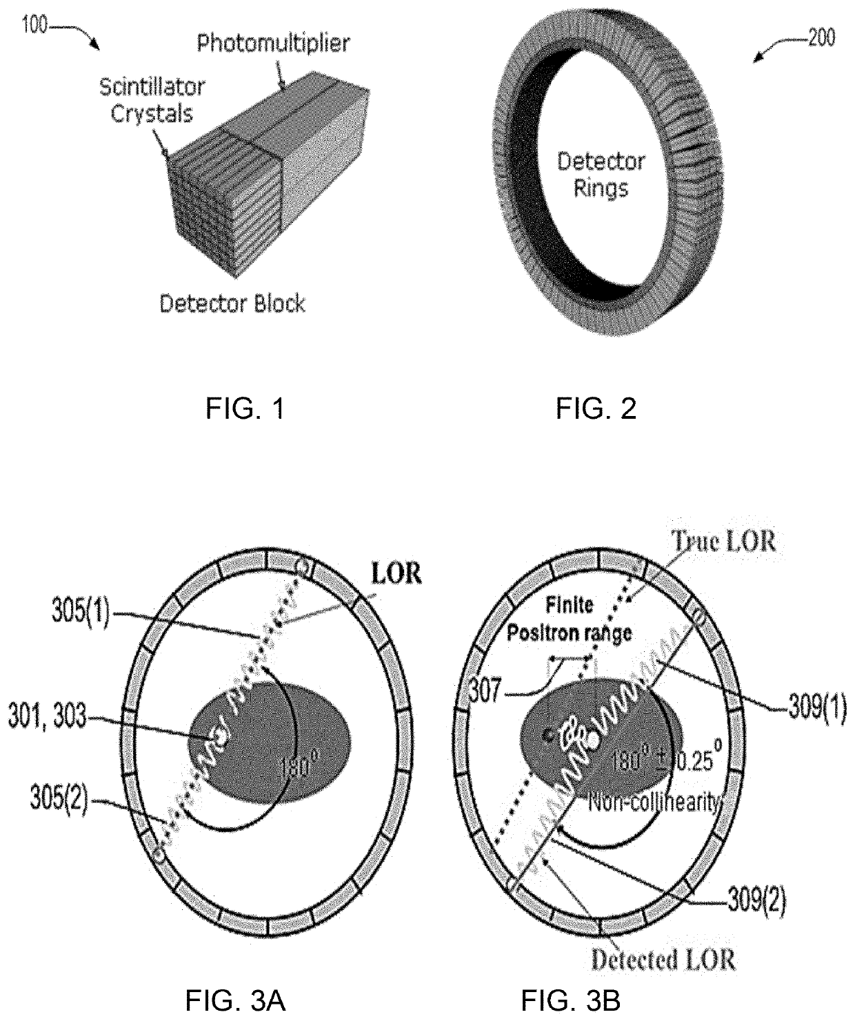
Figure 1 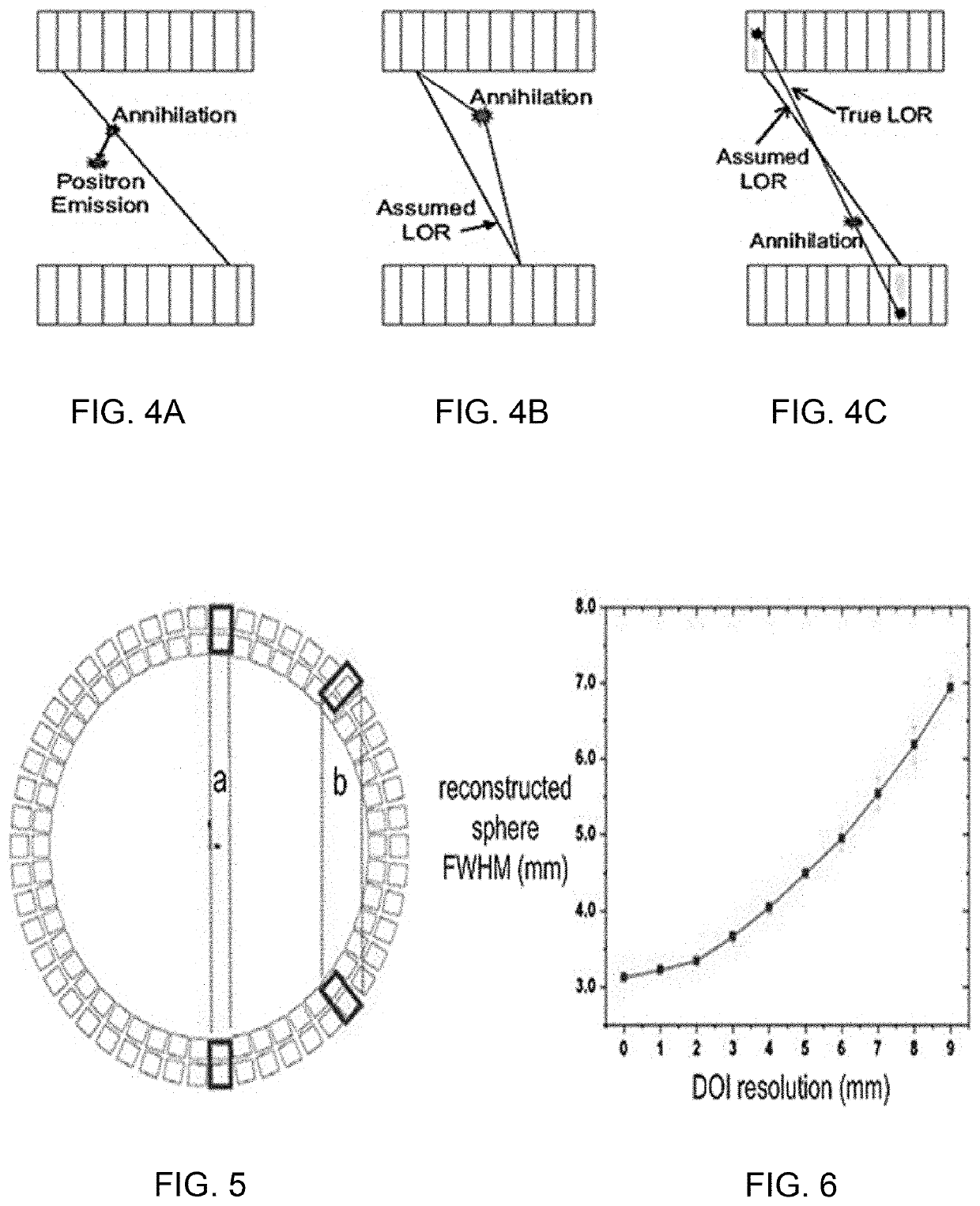
Figure 2 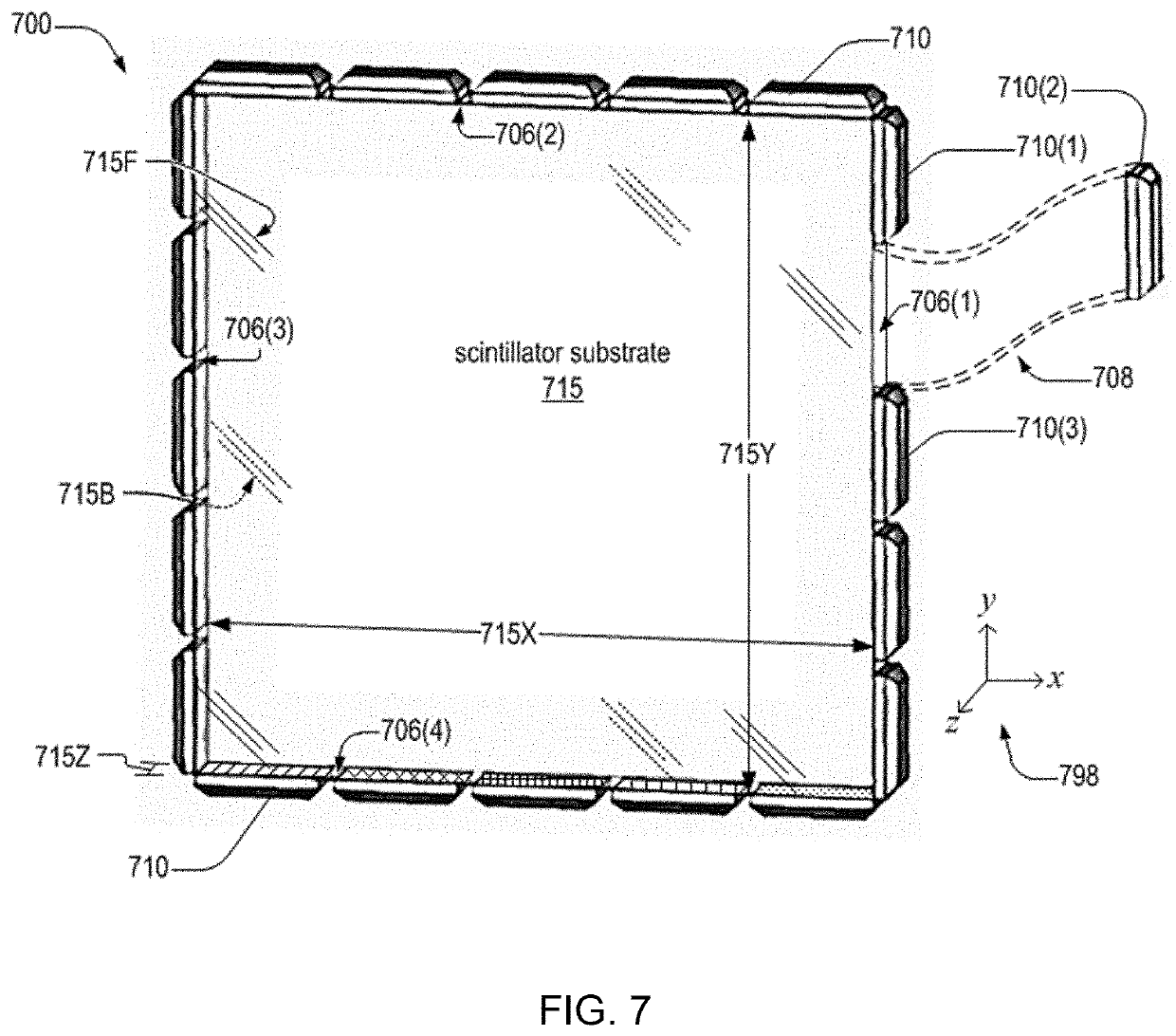
Figure 3
Abstract
Description
CROSS-REFERENCE TO RELATED APPLICATIONS
[0001] The present US patent application is a continuation-in-part from the U.S. patent application Ser. No. 16 / 345,682 filed on Apr. 26, 2019 and now published as US 2019 / 0353807, which is a national phase from the International Patent Application No. PCT / US17 / 58501 filed on Oct. 26, 2017 and now published as WO 2018 / 081404, which in turn claims priority from the U.S. Provisional Patent Application No. 62 / 414,469 filed on Oct. 28, 2016. The disclosure of each of the above-identified patent documents is incorporated herein by reference.STATEMENT REGARDING FEDERALLY SPONSORED RESEARCH OR DEVELOPMENT
[0002] This invention was made with government support under Grant No. P41 EB002035, awarded by NIH. The government has certain rights in the invention.BACKGROUND OF THE INVENTION
[0003] Positron Emission Tomography (PET) has become a mature and reliable technology. It has broad applications in neurology and oncology due to its ability to monitor metaboli...
Examples
example 1
[0094]An analysis of side readouts of monolithic scintillation crystals. A method of using the side surfaces of a thin monolithic scintillation crystal for reading out scintillation photons is described. A Monte-Carlo simulation was carried out for an LYSO crystal of 50.8 mm×50.8 mm×3 mm with five silicon photomultipliers attached on each of the four side surfaces. With 511 keV gamma-rays, X-Y spatial resolution of 2.10 mm was predicted with an energy resolution of 9%. The addition of optical barriers was also explored to improve the X-Y spatial resolution. An X-Y spatial resolution of 786 μm was predicted with an energy resolution of 9.2%. Multiple layers may be stacked together and readout channels may be combined. Depth of interaction information (DOI) may be directly read out. This method provides an attractive detector module design for positron emission tomography (PET).
[0095]FIG. 13 illustrates a scintillation event 1301 (or equivalently, a gamma-ray interaction event) within...
example 2
[0126]Simulation of a CsI / LYSO Detector Layer. This section describes simulation of crystals that that were tested experimentally. The geometry of the scintillation crystal was set as 27.4 mm×27.4 mm×3 mm. Sixteen SiPMs were attached to the edges of the scintillation crystal with 4 SiPMs on each edge. Several factors influencing the spatial resolution were studied:[0127]gamma-ray photon energy and scintillator material (662 keV for CsI(Tl) vs 511 keV for LYSO);[0128]optical barriers (with / without);[0129]optical-gel & SiPM-window refractive indices (1.5 vs 1.82).
[0130]The dark-count rate was set to 2 MHz / SiPM in accordance with the Hamamatsu S13360-6050PE MPPC data sheet. The readout shaping time was set to 4 μs for CsI(Tl) and 150 ns for LYSO, based on their published decay times (Mao R, Zhang L, Zhu, R. Optical and Scintillation Properties of Inorganic Scintillators in High Energy Physics. IEEE Transactions on Nuclear Science. 2008; 55(4): 2425-2431). The optical-gel / SiPM-window ma...
experimental verification
[0132]FIG. 40 includes photographs of components of a prototype detector 4001 and 4002. The prototype detector includes the following components, labelled (a) through (e) in FIG. 40:[0133](a) Sixteen Hamamatsu S13360-6050PE MPPCs (SiPMs);[0134](b) CsI(Tl) scintillation crystal: CsI(Tl) crystal of dimension 27.4 mm×27.4 mm×3 mm (obtained from Proteus, without mechanical holes as in detector 4001, and with mechanical holes, as in detector 4002);[0135](c) pre-amplifier circuit;[0136](d) amplifier and analog-to-digital-converting circuit;[0137](e) FPGA data acquisition circuit (AiT 16-channel-readout circuit).[0138]The photograph labelled (f) is prototype detector 4001 or 4002.
[0139]The scintillator was chosen to be CsI(Tl) for convenience (no background activity from the crystal and low hygroscopicity). The MDRFs were acquired by scanning the detector with a crossed-slit-collimated beam of 662 keV photons from a 137Cs source. The beam size on the detector surface was measured to be abo...