
Method for identifying compounds which affect ionotropic glutamate receptor aggregation
a technology of ionotropic glutamate receptor and compound, which is applied in the field of molecules involved in synaptogenesis, can solve the problems of additional complexity in the formation of central excitatory synapses, and achieve the effects of increasing the number of excitatory synapses
Patent Information
- Authority / Receiving Office
- US · United States
- Patent Type
- Patents(United States)
- Current Assignee / Owner
- Publication Date
- 2007-05-15
- Estimated Expiration
- Not applicable · inactive patent
Smart Images
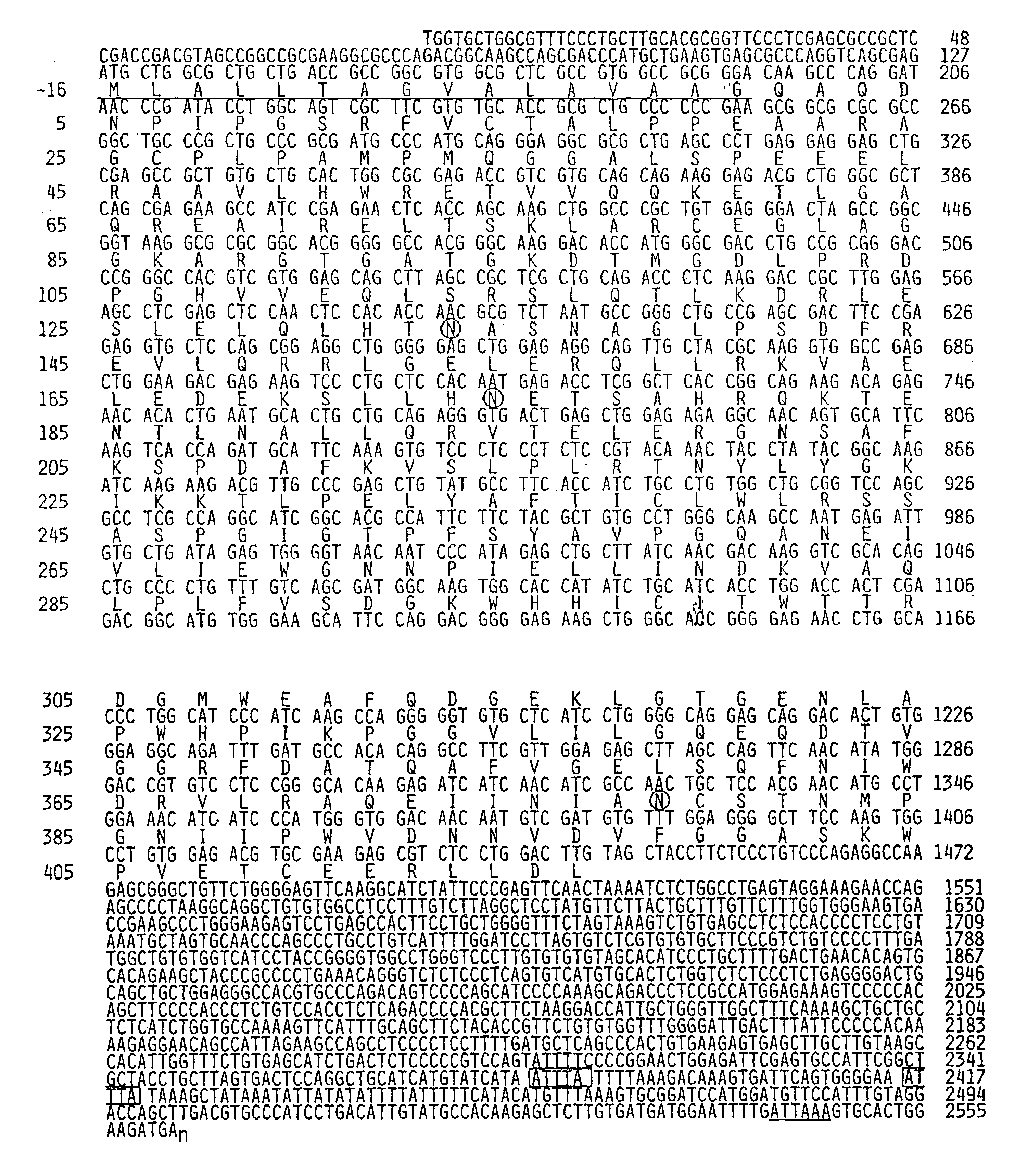
Figure 1 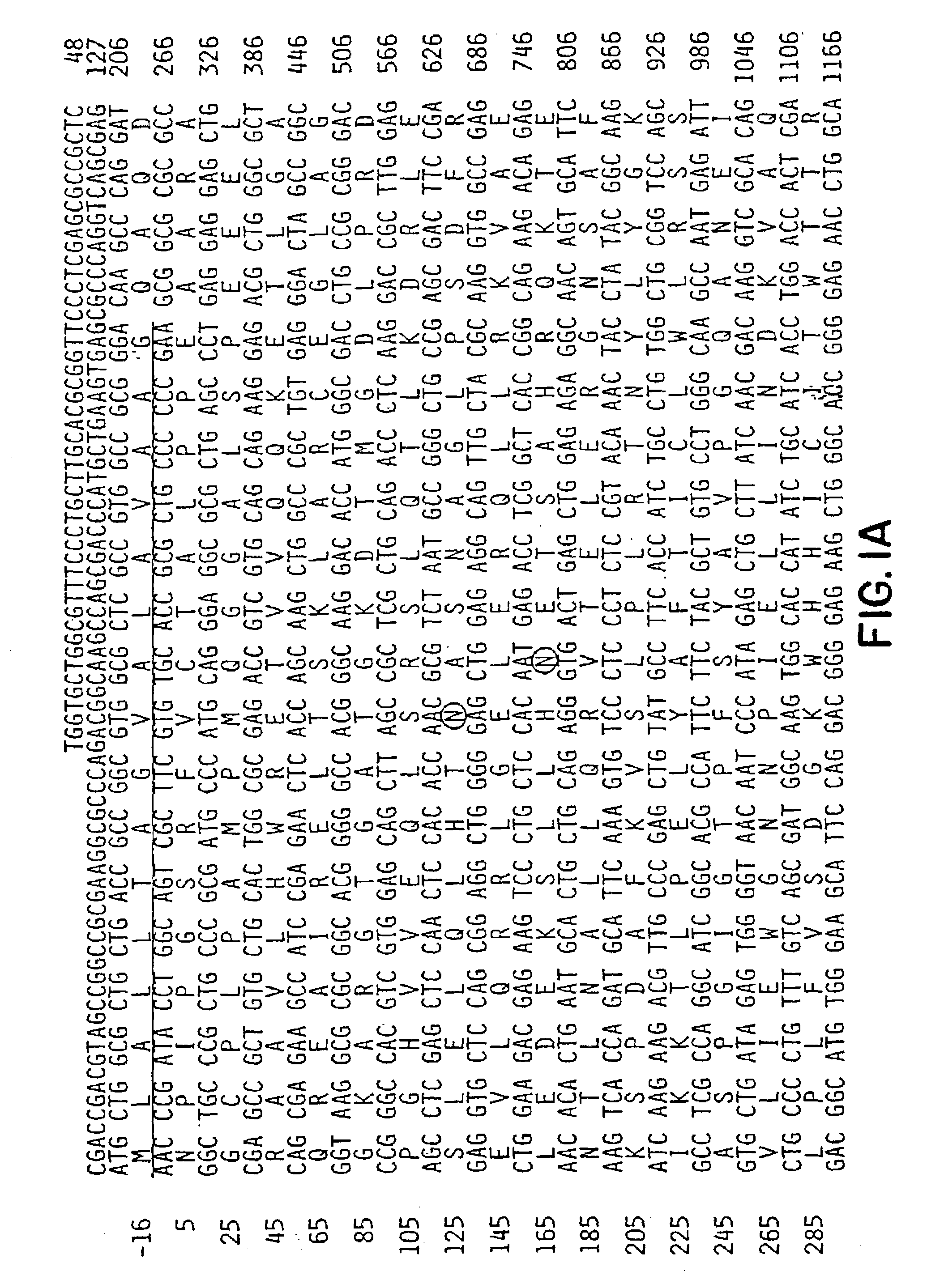
Figure 2 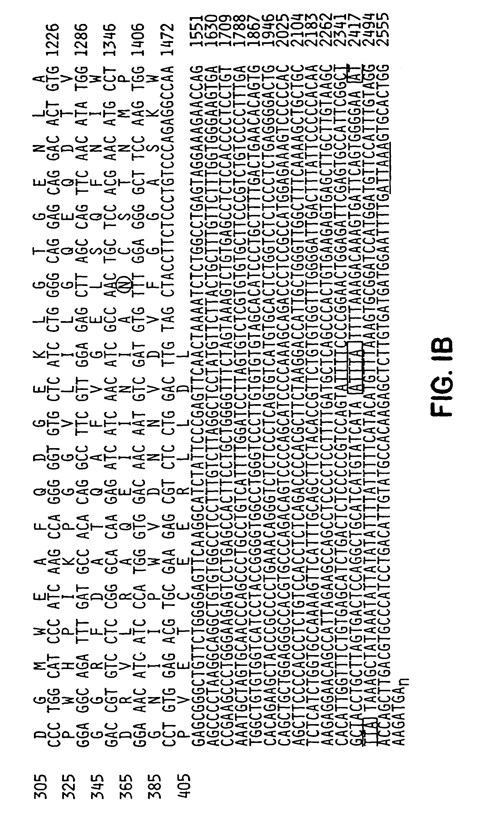
Figure 3
Abstract
Description
[0001] This is a continuation of U.S. patent application Ser. No. 09 / 328,710 filed Jun. 9, 1999, now abandoned, all of which is hereby incorporated by reference in the disclosure of this application.STATEMENT AS TO FEDERALLY SPONSORED RESEARCH
[0002] This invention was made with Government support under Grant No. K08NS01652, R01NS36715, R01MH53608, and K02MHO1152, awarded by the National Institutes of Health. The government may have certain rights in the invention.FIELD OF THE INVENTION
[0003] The present invention relates generally to molecules involved in synaptogenesis, and more specifically to the role of Narp in the formation of synapses and the clustering of receptors in synapses.BACKGROUND OF THE INVENTION
[0004] The mature central nervous system exhibits the capacity to alter cellular interactions as a function of the activity of specific neuronal circuits. This capacity is believed to underlie learning and memory storage, age-related memory loss, tolerance to and dependence on dru...
Examples
example 1
NARP is Enriched at Excitatory Synapses in a Subpopulation of Neurons from Hippocampus and Spinal Cord
[0087]To study Narp protein, a rabbit polyclonal antibody was generated against a full length GST fusion protein of Narp. On western blot, this antibody recognized a single broad protein band centered at 56 kDa in rat brain, similar to the size of recombinant Narp expressed in detergent extracts of transfected HEK-293 cells. Narp protein was also detected as a similar sized triplet in rat testes, but not in other peripheral tissues. This restricted distribution of Narp protein parallels the Narp mRNA expression previously reported (Tsui et al., supra, 1996). The broadness of the Narp band in brain is consistent with the observation that Narp is glycosylated (Tsui et al., supra, 1996). In non-reducing gels, Narp migrates as a multimer, with a size greater than 220 kDa, consistent with the known ability of members of the pentraxin family to covalently multimerize through disulfide bon...
example 2
Transfected Narp Accumulates at Excitatory Synapses
[0094]In order to characterize the synaptic targeting of Narp in neurons, a C-terminal myc-tagged version of Narp (designated myc-Narp) was transfected into cultured spinal neurons (Dong et al., Nature 386:279–84, 1997). After 72 hours of expression, live staining with an anti-myc (mouse) antibody, followed by a FITC labeled anti-mouse secondary, was used to reveal the surface distribution of the transfected Narp. Subsequent staining of the same cells, after fixation and permeabilization, with an AMCA labeled anti-myc (rabbit) antibody was used to identify transfected neurons and their axons and dendrites, while GluR1 and GAD staining were used to identify excitatory and inhibitory synapses, respectively.
[0095]Staining of transfected neurons indicated that myc-Narp, similar to endogenous Narp, was distributed throughout the somato-dendritic domain of the transfected neuron and was also specifically localized to excitatory synapses. ...
example 3
Synaptic Narp is Derived from Both the Presynaptic and Postsynaptic Neuron
[0097]Since only 1 to 3% of the neurons in a given experiment are transfected by the procedure described above, it was relatively easy to distinguish myc-Narp that came from the postsynaptic cell (surface stain associated with the large proximal processes of an isolated transfected cell) from myc-Narp originating from the presynaptic cell (surface stain associated with a solitary thin process coursing over a non-transfected cell body or dendrite which is not in continuity with any visibly transfected cell). This operationally defined distinction between axon and dendrite was verified to be nearly 90% specific in pilot experiments in which labeled processes were stained with anti-Tau antibodies to distinguish axons from dendrites. As discussed above, 74% of excitatory synapses in transfected neurons had clear surface myc-Narp stain, indicating that Narp is targeted to the synapse from the postsynaptic neuron. H...