
Method for regenerating cartilage
a cartilage composition and regenerative technology, applied in the field of methods, can solve the problems of enlargement and progression of lesion, limited repair potential of articular cartilage of higher animals, and enlargement of lesion, and achieve the effect of effective cartilage composition and reliable inducement of new cartilage formation
Patent Information
- Authority / Receiving Office
- US · United States
- Patent Type
- Patents(United States)
- Current Assignee / Owner
- Publication Date
- 2010-11-23
Smart Images
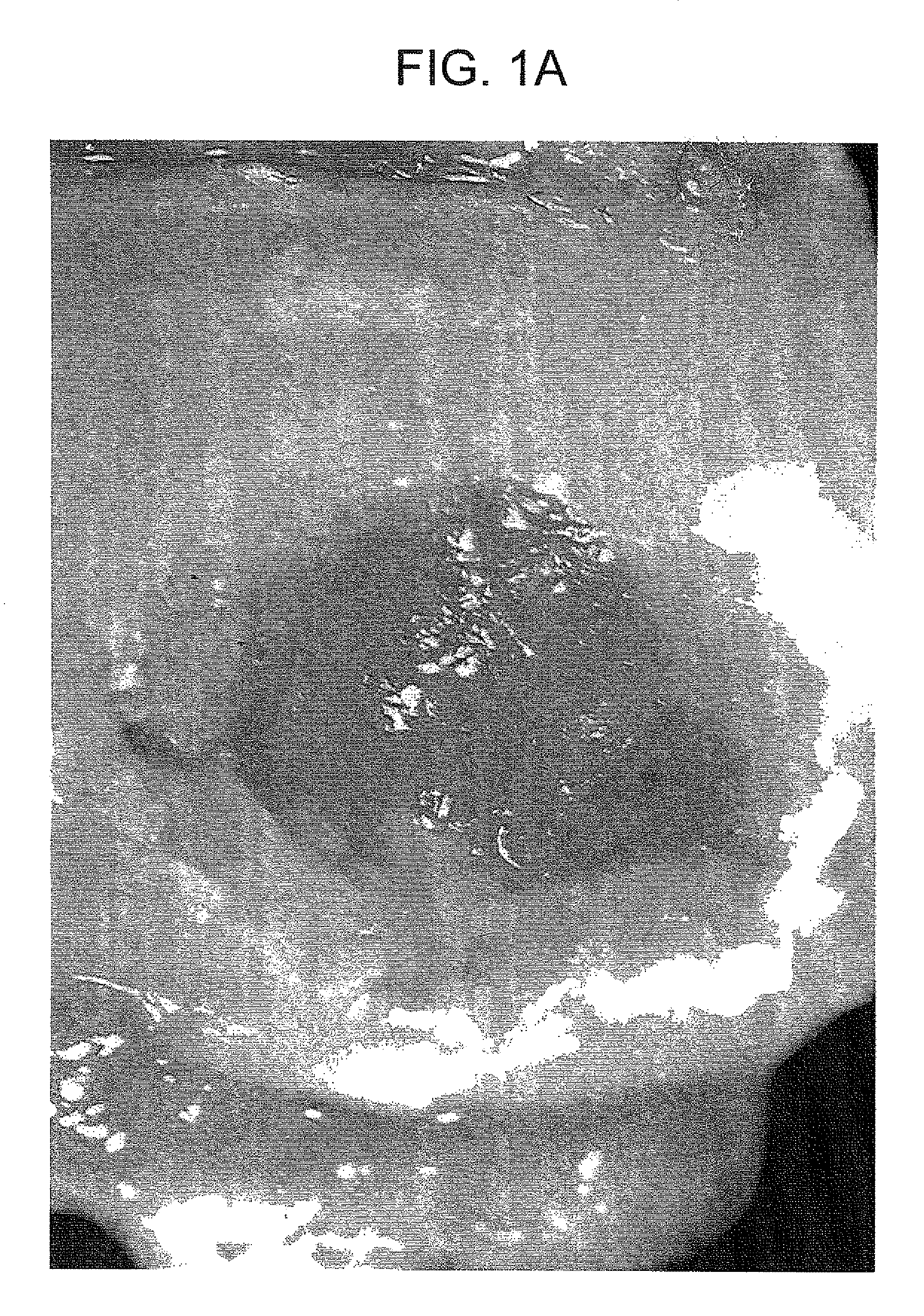
Figure 1 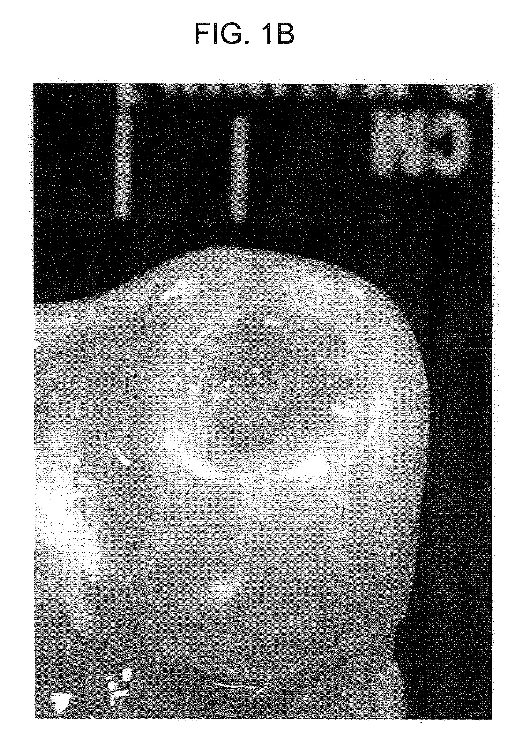
Figure 2 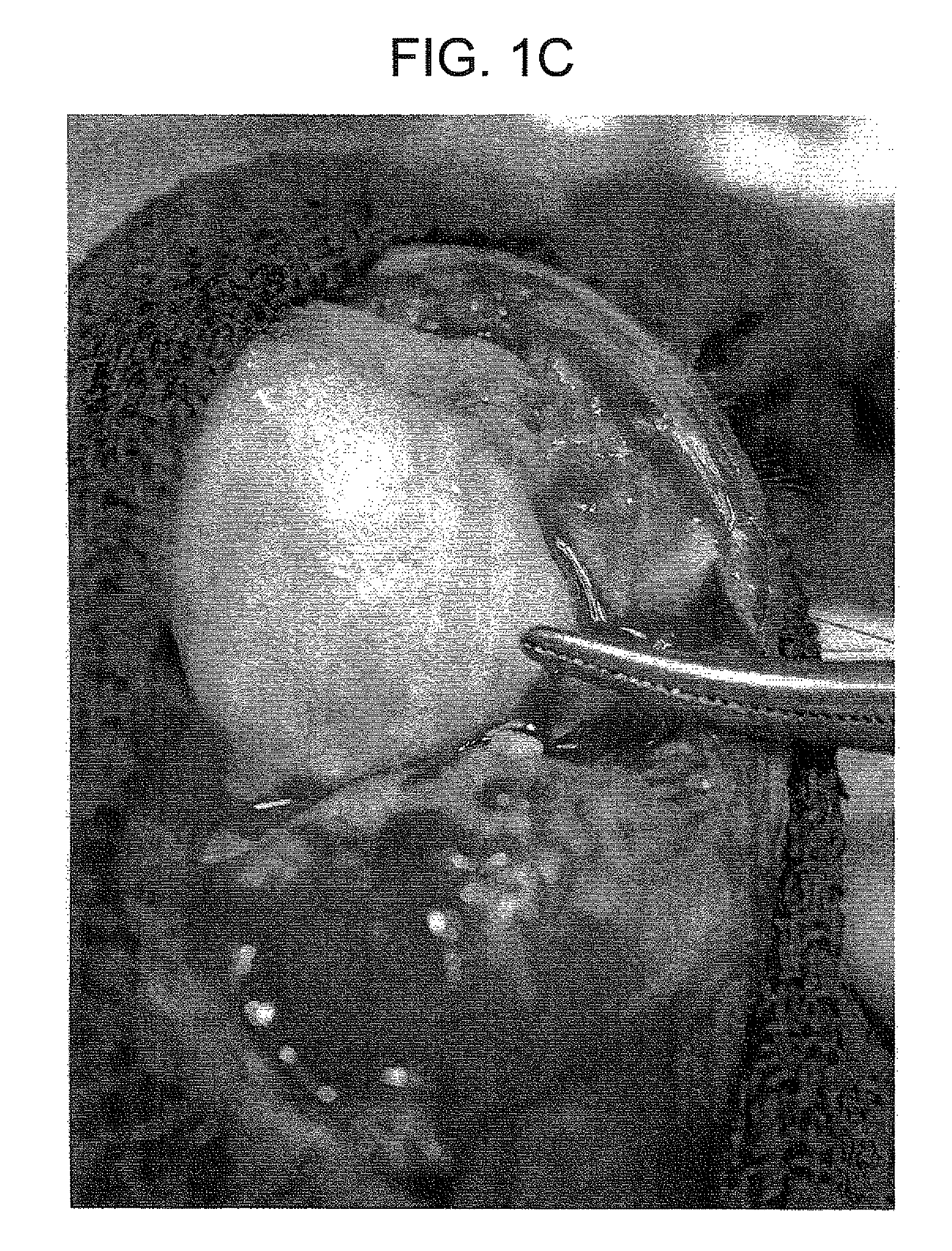
Figure 3
Abstract
Description
CROSS-REFERENCE TO A RELATED APPLICATION
[0001] This patent application is a divisional of copending U.S. patent application Ser. No. 10 / 793,939, filed Mar. 8, 2004, the disclosure of which is incorporated by reference.FIELD OF THE INVENTION
[0002] This invention pertains to particulate cartilage compositions, processes for preparing particulate cartilage compositions and methods for regenerating articular cartilage.BACKGROUND OF THE INVENTION
[0003] Articular cartilage of higher animals, including man, has very limited potential for repair. Following injuries to articular cartilage, the cartilage generally does not repair itself because of its very limited capacity for regeneration. Repair is dependent on the extent and the depth of injury and the surviving chondrocytes and normal articular cartilage matrix. In injuries involving subchondral bone, there is no regeneration of the cartilage, but there is typically enlargement and progression of the lesion with associated pain. The ultimate...
Examples
example
[0031]This example demonstrates the chondrogenic activity of the inventive composition in fully immunocompetant non-human primates. Full thickness cartilage defects measuring 10 min×10 mm were created in the medial condyles of the animals. The defects were densely packed with the cartilage composition according to the present invention and were compacted with a tamp. The animals were examined two, six, and sixteen weeks post transplantation and the joints were re-explored. FIGS. 1a-1c are photographs taken at two, six, and sixteen weeks, respectively. Specimens were also taken and were fixed in 10% formalin-Earle's balanced salt solutions. Paraffin sections were cut and stained with homotoxylin and eosin, PAS, Romanowski-Giemsa and Safranin-O stains. FIGS. 2a and 2b illustrate the specimens taken (100×) at six and sixteen weeks, respectively.
[0032]As seen in FIG. 1a, at two weeks post transplantation, granulation tissue is present in the center of the defect and new cartilage is pre...