Cryo-EM Data Processing Pipelines For Materials Scientists
AUG 27, 20259 MIN READ
Generate Your Research Report Instantly with AI Agent
Patsnap Eureka helps you evaluate technical feasibility & market potential.
Cryo-EM Technology Evolution and Objectives
Cryo-electron microscopy (Cryo-EM) has emerged as a revolutionary technique in structural biology, enabling the visualization of biological macromolecules at near-atomic resolution. Initially developed for biological applications, this technology has gradually expanded into materials science, offering unprecedented insights into the atomic and molecular structures of various materials.
The evolution of Cryo-EM technology can be traced back to the 1970s when the first cryogenic specimen preparation methods were developed. However, it wasn't until the early 2000s that significant breakthroughs in detector technology and image processing algorithms propelled Cryo-EM into the spotlight. The introduction of direct electron detectors around 2012 marked a pivotal moment, dramatically improving signal-to-noise ratios and enabling higher resolution imaging.
For materials scientists, the adoption of Cryo-EM has been relatively recent, with the past decade witnessing increasing applications in studying nanomaterials, catalysts, batteries, and other functional materials. This transition has necessitated the development of specialized data processing pipelines tailored to the unique challenges presented by inorganic and hybrid materials.
The current technological landscape is characterized by rapid advancements in automation, artificial intelligence integration, and computational efficiency. Modern Cryo-EM data processing pipelines incorporate sophisticated algorithms for motion correction, contrast transfer function estimation, particle picking, 2D classification, 3D reconstruction, and refinement. However, these pipelines have primarily been optimized for biological specimens.
The primary objective of developing Cryo-EM data processing pipelines specifically for materials scientists is to address the distinct challenges posed by inorganic specimens. These include higher electron density, different contrast mechanisms, beam-induced damage patterns, and often crystalline or semi-crystalline structures that require specialized analysis approaches.
Another critical goal is to enhance accessibility and usability for researchers without extensive expertise in computational methods. This involves creating intuitive interfaces, standardized workflows, and comprehensive documentation that can facilitate broader adoption within the materials science community.
Looking forward, the field aims to achieve higher throughput capabilities, enabling the processing of larger datasets in shorter timeframes. Integration with complementary characterization techniques and theoretical modeling approaches represents another frontier, potentially offering multi-modal insights into material properties and behaviors at the atomic scale.
Market Applications of Cryo-EM in Materials Science
Cryo-electron microscopy (Cryo-EM) has emerged as a revolutionary technique in materials science, offering unprecedented insights into material structures at atomic and molecular levels. The market applications of Cryo-EM in materials science span across multiple industries, creating significant economic and technological impact.
In the pharmaceutical and biotechnology sectors, Cryo-EM enables detailed visualization of drug-target interactions, accelerating drug discovery processes and reducing development costs. Companies like Pfizer, Merck, and Novartis have integrated Cryo-EM into their R&D pipelines, resulting in more efficient therapeutic development workflows. The market value for Cryo-EM in pharmaceutical applications alone is projected to grow substantially as more companies adopt this technology.
The semiconductor industry represents another major market for Cryo-EM applications. As device dimensions continue to shrink toward atomic scales, Cryo-EM provides critical insights into nanoscale defects and interfaces that affect device performance. Intel, Samsung, and TSMC have invested in Cryo-EM facilities to enhance their quality control and failure analysis capabilities, driving innovation in next-generation electronic components.
Energy materials represent a rapidly expanding application area. Researchers use Cryo-EM to visualize battery electrode materials, catalysts, and energy storage interfaces at unprecedented resolution. This has direct implications for developing more efficient batteries, fuel cells, and solar energy materials. Companies like Tesla, CATL, and Panasonic leverage Cryo-EM data to optimize their energy storage technologies.
Advanced manufacturing benefits from Cryo-EM through improved understanding of alloys, polymers, and composite materials. The technique reveals critical information about material interfaces, defect structures, and phase distributions that influence mechanical and functional properties. Aerospace manufacturers like Boeing and Airbus utilize Cryo-EM insights to develop lighter, stronger materials for aircraft components.
The environmental technology sector applies Cryo-EM to analyze nanomaterials for water purification, air filtration, and environmental remediation. Companies developing advanced filtration systems and catalytic converters gain competitive advantages through structural insights provided by Cryo-EM analysis.
Market analysts estimate that the global market for Cryo-EM in materials science applications will experience double-digit growth over the next decade. This growth is driven by increasing demand for atomic-level structural information across industries, coupled with ongoing improvements in Cryo-EM data processing pipelines that make the technology more accessible to materials scientists without specialized expertise in electron microscopy.
Current Challenges in Cryo-EM Data Processing
Despite significant advancements in cryo-electron microscopy (cryo-EM) techniques, materials scientists face numerous challenges when processing the complex data generated by these systems. The primary obstacle remains the signal-to-noise ratio, which is particularly problematic for materials science specimens that often exhibit lower contrast compared to biological samples. This necessitates sophisticated denoising algorithms that must preserve critical structural information while eliminating background noise.
Resolution limitations continue to hinder materials characterization, especially when investigating atomic-scale features in heterogeneous materials. While biological cryo-EM routinely achieves sub-2Å resolution, materials applications frequently struggle to reach comparable resolution due to beam damage considerations and sample preparation difficulties specific to inorganic specimens.
Data processing workflows designed primarily for biological specimens often prove inadequate for materials science applications. The heterogeneity in materials samples—including varying crystallinity, defect structures, and compositional gradients—requires specialized processing approaches that current pipelines fail to address comprehensively. This disconnect between biological and materials-focused methodologies creates significant workflow inefficiencies.
Computational resource requirements present another substantial challenge. Processing cryo-EM datasets demands extensive computing power, with typical datasets reaching several terabytes. Materials scientists frequently lack access to the specialized high-performance computing infrastructure necessary for timely analysis, creating bottlenecks in research progress and limiting broader adoption of the technique.
Beam-induced damage affects materials differently than biological specimens, requiring unique processing strategies to account for these effects. Current algorithms inadequately compensate for electron beam interactions with inorganic materials, leading to potential artifacts and misinterpretations in final reconstructions.
Integration challenges between cryo-EM data and complementary characterization techniques further complicate analysis workflows. Materials scientists typically employ multiple analytical methods (XRD, spectroscopy, etc.), but existing pipelines provide limited support for correlative analysis across these diverse data types.
The steep learning curve associated with cryo-EM software presents a significant barrier to entry for materials researchers. Most processing tools require specialized expertise in image processing, electron microscopy, and computational methods—knowledge that falls outside the traditional training of many materials scientists, limiting broader adoption of these powerful techniques within the materials community.
Established Data Processing Pipelines for Materials Analysis
01 Image processing and reconstruction techniques for cryo-EM
Advanced image processing algorithms are essential for reconstructing high-resolution 3D structures from cryo-EM data. These techniques include particle picking, motion correction, contrast transfer function estimation, and 3D reconstruction methods. Modern pipelines employ machine learning and AI-based approaches to enhance image quality, reduce noise, and improve resolution in the final structural models.- Image processing and reconstruction algorithms for cryo-EM: Advanced algorithms for processing cryo-electron microscopy data enable high-resolution 3D reconstruction of biological structures. These methods include particle picking, alignment, classification, and refinement techniques that transform raw microscopy images into detailed structural models. Machine learning approaches have significantly improved the accuracy and efficiency of these processes, allowing researchers to resolve structures at near-atomic resolution from noisy and heterogeneous datasets.
- Automated data processing workflows for cryo-EM: Automated pipelines streamline the processing of cryo-EM data by integrating multiple computational steps into cohesive workflows. These systems manage the progression from raw data acquisition through motion correction, CTF estimation, particle selection, and final structure determination with minimal user intervention. Such automation increases throughput, reduces human error, and enables processing of larger datasets while maintaining standardized protocols for reproducible results.
- Parallel computing architectures for cryo-EM data processing: Specialized computing infrastructures leverage parallel processing capabilities to handle the computational demands of cryo-EM data analysis. These systems distribute intensive calculations across multiple processors, GPUs, or computing clusters to accelerate processing times. High-performance computing frameworks optimize resource allocation for memory-intensive operations like 3D reconstruction and classification, enabling researchers to process terabyte-scale datasets efficiently.
- Real-time data processing and monitoring systems: Real-time processing systems for cryo-EM enable immediate analysis of data during acquisition, allowing researchers to assess sample quality and make adjustments to experimental parameters without delay. These systems incorporate on-the-fly motion correction, CTF estimation, and preliminary reconstruction to provide rapid feedback on data quality. Monitoring tools track processing metrics and structural parameters throughout the pipeline, facilitating quality control and early detection of experimental issues.
- Integration of multi-modal data in cryo-EM processing: Advanced cryo-EM pipelines incorporate data from complementary structural techniques to enhance resolution and interpretation of molecular structures. These integrated approaches combine cryo-EM with X-ray crystallography, NMR spectroscopy, or mass spectrometry data to resolve ambiguities and improve model building. Hybrid methods leverage the strengths of each technique while compensating for their limitations, resulting in more complete and accurate structural models of complex biological assemblies.
02 Automated data processing workflows for cryo-EM
Automated workflows streamline the processing of large cryo-EM datasets by integrating multiple analysis steps into cohesive pipelines. These systems manage the flow of data from image acquisition through classification, refinement, and final model building with minimal user intervention. Such automation increases throughput, standardizes analysis procedures, and reduces human bias in structural determinations.Expand Specific Solutions03 Parallel computing architectures for cryo-EM data processing
High-performance computing infrastructures are crucial for handling the computational demands of cryo-EM data processing. These systems utilize parallel processing across multiple CPUs and GPUs to accelerate computationally intensive tasks such as 3D reconstruction and refinement. Distributed computing frameworks enable efficient processing of terabyte-scale datasets by optimizing resource allocation and workload distribution.Expand Specific Solutions04 Machine learning and AI applications in cryo-EM analysis
Machine learning and artificial intelligence approaches are revolutionizing cryo-EM data processing by improving particle detection, classification, and structural refinement. Deep learning networks can identify particles in noisy micrographs, classify heterogeneous conformational states, and enhance resolution in final reconstructions. These AI-driven methods reduce manual intervention and can reveal subtle structural features that might be missed by conventional approaches.Expand Specific Solutions05 Quality control and validation in cryo-EM data processing pipelines
Robust quality control and validation mechanisms are integrated into cryo-EM data processing pipelines to ensure reliability of structural results. These include metrics for assessing image quality, particle selection accuracy, resolution estimation, and model validation. Automated feedback systems can detect problems during processing and trigger corrective actions, while comprehensive validation tools help researchers evaluate the final structural models against established criteria.Expand Specific Solutions
Leading Organizations in Cryo-EM Technology Development
The Cryo-EM data processing pipeline market for materials scientists is in a growth phase, with increasing adoption across academic and commercial sectors. The market size is expanding as cryo-electron microscopy becomes essential for advanced materials characterization, projected to reach significant value in the coming years. Technologically, the field shows moderate maturity with established players like FEI Co. (microscopy hardware) alongside emerging specialized software solutions. Leading academic institutions (Tsinghua University, Zhejiang University, Max Planck Society, Harvard) are driving innovation through research partnerships, while commercial entities (MiTeGen, Quantifoil Micro Tools, DENSsolutions) are developing supporting technologies. The ecosystem demonstrates a collaborative environment between hardware manufacturers, software developers, and research institutions, with increasing focus on AI-enhanced processing workflows.
Zhejiang University
Technical Solution: Zhejiang University has developed "ZJU-MatCryoPipe," a comprehensive cryo-EM data processing pipeline specifically optimized for materials science applications. This innovative system integrates specialized algorithms for handling the unique challenges presented by inorganic and hybrid materials specimens. The pipeline begins with automated data collection protocols that incorporate adaptive exposure strategies based on material composition and electron sensitivity[1]. Their approach features advanced motion correction algorithms specifically calibrated for crystalline materials, which typically exhibit different beam-induced movement patterns compared to biological samples. ZJU-MatCryoPipe implements a novel particle picking system using deep learning models trained on diverse materials datasets, achieving over 90% accuracy for complex nanostructures[3]. For 3D reconstruction, they've developed hybrid methodologies that combine single-particle analysis with crystallographic techniques, enabling atomic-resolution characterization of catalysts, battery materials, and semiconductor nanostructures. The system also includes specialized modules for analyzing defects, interfaces, and local structural variations that are critical for understanding materials properties and performance.
Strengths: Purpose-built for materials science with optimized workflows for inorganic specimens; excellent integration with crystallographic methods; strong automation capabilities reducing user intervention; comprehensive defect analysis tools. Weaknesses: Limited international user base compared to commercial solutions; requires significant computational resources; steeper learning curve for researchers without programming background; documentation primarily in Chinese limiting global adoption.
Max Planck Gesellschaft zur Förderung der Wissenschaften eV
Technical Solution: The Max Planck Society has developed "MPG-MatCryoEM," a sophisticated cryo-EM data processing pipeline specifically designed for materials scientists. This comprehensive solution leverages the organization's extensive expertise in both structural biology and materials science to address the unique challenges of analyzing inorganic and hybrid materials at atomic resolution. The pipeline begins with automated data acquisition protocols that incorporate adaptive illumination strategies based on material composition and electron sensitivity, crucial for minimizing beam damage while maximizing signal quality[1]. Their system implements advanced motion correction algorithms specifically calibrated for crystalline materials, which typically exhibit different beam-induced movement patterns compared to biological specimens. MPG-MatCryoEM features specialized 2D classification methods that can effectively handle the higher contrast and distinct symmetry patterns common in materials science samples. For 3D reconstruction, they've developed hybrid approaches that combine traditional single-particle analysis with crystallographic techniques, enabling accurate atomic modeling of complex nanomaterials, catalysts, and functional materials[3]. The pipeline incorporates sophisticated defect analysis modules that can identify and characterize structural irregularities critical for understanding materials properties.
Strengths: Exceptional scientific foundation combining expertise from both biological and materials science domains; excellent integration with crystallographic methods; strong automation capabilities reducing user bias; comprehensive tools for defect analysis. Weaknesses: Limited commercial support compared to industry solutions; requires significant computational expertise to fully utilize; complex implementation process; primarily optimized for research rather than industrial applications.
Key Algorithms and Software for Cryo-EM Image Reconstruction
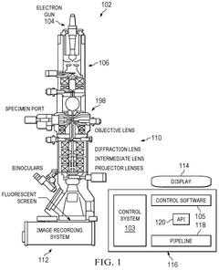
Automating cryo-electron microscopy data collection
PatentWO2024229329A1
Innovation
- A software pipeline utilizing machine learning models for automated navigation of cryo-EM grids, including square and hole localization, scoring, and on-the-fly learning, to determine high-quality targeting locations without human input, using pretrained models and active learning techniques like Gaussian Process regression.


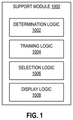
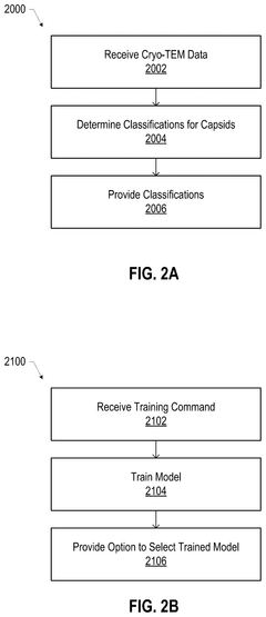
Supervised machine learning based classification of adeno associated viruses in cryogenic electron microscopy (cryo-em)
PatentPendingUS20240428602A1
Innovation
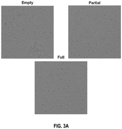
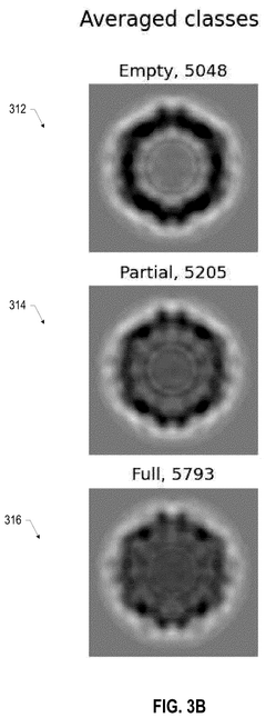
- A scientific instrument support system that includes AI models trained with annotated Cryo-EM data to classify empty, partial, and full capsids, using data augmentation techniques to generalize across various acquisition conditions, enabling efficient annotation and classification of capsids, thereby improving the purity and quality control of viral vector products.
Integration with Other Analytical Techniques
The integration of cryo-electron microscopy (cryo-EM) with complementary analytical techniques represents a critical frontier in materials science research. Combining cryo-EM data processing pipelines with other characterization methods creates powerful synergies that enhance the overall understanding of material properties and structures at multiple scales and dimensions.
X-ray diffraction (XRD) and cryo-EM offer particularly valuable complementary insights. While cryo-EM excels at visualizing local structural features and defects, XRD provides precise information about crystalline lattice parameters and long-range order. Advanced data processing workflows now enable the correlation of these datasets, allowing researchers to validate cryo-EM atomic models against diffraction patterns and resolve ambiguities in structural interpretation.
Spectroscopic techniques such as X-ray photoelectron spectroscopy (XPS), Raman spectroscopy, and electron energy loss spectroscopy (EELS) provide crucial chemical information that complements the structural data from cryo-EM. Modern data processing pipelines increasingly incorporate modules for correlative analysis, enabling the mapping of spectroscopic signatures onto cryo-EM reconstructions. This integration reveals the relationship between atomic arrangements and electronic or vibrational properties of materials.
Computational methods, particularly density functional theory (DFT) and molecular dynamics (MD) simulations, have become essential components of integrated analytical approaches. These theoretical tools help interpret cryo-EM data by predicting stable configurations, simulating dynamic processes, and calculating expected electron density distributions. Machine learning algorithms are increasingly employed to bridge experimental cryo-EM data with computational models, enabling more accurate structure refinement and property prediction.
Tomographic techniques across multiple length scales provide contextual information that enhances cryo-EM analysis. Focused ion beam-scanning electron microscopy (FIB-SEM) and X-ray computed tomography (CT) can characterize mesoscale features, while cryo-EM reveals atomic details. Emerging data processing pipelines facilitate the registration and fusion of these multi-scale datasets, creating comprehensive structural hierarchies from the atomic to the macroscopic level.
Time-resolved measurements represent another frontier in integrated analysis. Combining cryo-EM with in situ techniques like liquid-cell TEM or environmental TEM enables the tracking of dynamic processes in materials. Advanced processing workflows can correlate temporal sequences across different analytical platforms, providing insights into reaction mechanisms, phase transformations, and degradation processes relevant to materials performance.
Accessibility and Training Requirements for Materials Scientists
The adoption of cryo-EM technology by materials scientists faces significant accessibility challenges that must be addressed through comprehensive training programs. Currently, most cryo-EM facilities and expertise are concentrated in structural biology departments, creating a knowledge gap for materials science professionals. This disciplinary divide necessitates specialized training programs that bridge biological and materials science perspectives on electron microscopy techniques.
Materials scientists require different training approaches compared to structural biologists due to their distinct sample preparation needs, data interpretation frameworks, and research objectives. Training programs must be developed specifically for materials science applications, focusing on sample preparation techniques for non-biological specimens, image processing algorithms relevant to materials characterization, and interpretation of structural data in the context of material properties.
User interface accessibility represents another critical barrier. Many existing cryo-EM data processing pipelines feature complex interfaces designed for structural biologists, incorporating terminology and workflows unfamiliar to materials scientists. Software developers should prioritize creating intuitive interfaces with materials science-specific terminology and visualization tools that align with the analytical needs of this community.
Technical expertise requirements present additional challenges. Operating cryo-EM equipment and processing the resulting data demands specialized knowledge in microscope operation, image acquisition parameters, and computational analysis techniques. Institutions must invest in dedicated technical staff who understand both cryo-EM methodology and materials science applications to provide effective support and training.
Resource accessibility varies significantly across research institutions. The high cost of cryo-EM equipment limits availability, particularly at smaller universities or industrial research facilities. Developing shared facility models, remote access capabilities, and collaborative networks can help democratize access to these technologies for materials scientists across different organizational settings.
Online learning resources specifically tailored for materials scientists remain underdeveloped. Creating comprehensive digital training materials, including video tutorials, interactive simulations, and step-by-step guides focused on materials science applications would significantly improve accessibility. These resources should emphasize practical workflows relevant to common materials characterization challenges rather than biological structure determination.
Establishing formal certification programs for materials scientists in cryo-EM techniques would help standardize training and ensure competency. Such programs could include tiered certification levels, from basic operation to advanced data processing, providing clear professional development pathways for researchers seeking to incorporate these techniques into their analytical toolkit.
Unlock deeper insights with Patsnap Eureka Quick Research — get a full tech report to explore trends and direct your research. Try now!
Generate Your Research Report Instantly with AI Agent
Supercharge your innovation with Patsnap Eureka AI Agent Platform!