

Cryo-EM For 2D Material Heterostructure Interface Analysis
AUG 27, 20259 MIN READ
Generate Your Research Report Instantly with AI Agent
PatSnap Eureka helps you evaluate technical feasibility & market potential.
Cryo-EM Technology Evolution and Research Objectives
Cryo-electron microscopy (Cryo-EM) has emerged as a revolutionary technique in the field of structural biology, enabling researchers to visualize biological macromolecules at near-atomic resolution. In recent years, this technology has been increasingly adapted for materials science applications, particularly for the analysis of 2D material heterostructures and their interfaces. The evolution of Cryo-EM technology represents a convergence of advances in electron optics, detector technology, and computational methods.
The journey of Cryo-EM began in the 1970s with the development of vitrification techniques by Jacques Dubochet, who later received the Nobel Prize in Chemistry in 2017 alongside Richard Henderson and Joachim Frank. The breakthrough allowed biological samples to be preserved in their native state without the formation of ice crystals that could damage the specimen structure. By the early 2000s, the resolution capabilities had improved significantly, but were still limited compared to X-ray crystallography.
A pivotal moment in Cryo-EM evolution came with the introduction of direct electron detectors around 2012-2013, which dramatically improved signal-to-noise ratios and enabled the collection of movie frames rather than single images. This advancement, coupled with sophisticated image processing algorithms, led to the "resolution revolution," pushing the boundaries of what could be visualized at the atomic level.
For 2D material heterostructures, which consist of layers of different 2D materials stacked together, understanding the interface properties is crucial as they often determine the overall performance of devices. Traditional characterization techniques like transmission electron microscopy (TEM) have limitations in preserving the pristine state of these interfaces due to sample preparation requirements and beam damage effects.
The application of Cryo-EM to 2D material heterostructures aims to overcome these limitations by maintaining the samples at cryogenic temperatures, thereby minimizing beam damage and preserving the native structure of interfaces. This approach enables researchers to study the atomic arrangements, bonding characteristics, and electronic properties at heterointerfaces with unprecedented clarity.
Current research objectives in this field include developing specialized sample preparation techniques for 2D materials, optimizing imaging parameters for non-biological specimens, and enhancing computational methods for extracting structural information from low-contrast images. Additionally, there is a growing interest in combining Cryo-EM with spectroscopic techniques to correlate structural features with electronic and optical properties.
The ultimate goal is to establish Cryo-EM as a standard tool for comprehensive characterization of 2D material heterostructures, providing insights that can guide the rational design of next-generation electronic, optoelectronic, and energy storage devices. This would represent a significant step forward in bridging the gap between fundamental materials science and practical applications in various technological domains.
Market Applications for 2D Material Interface Analysis
The integration of Cryo-EM technology for 2D material heterostructure interface analysis presents significant market opportunities across multiple industries. The semiconductor industry stands as a primary beneficiary, where precise interface characterization enables the development of next-generation electronic devices with enhanced performance characteristics. Manufacturers of integrated circuits and transistors are increasingly adopting 2D materials like graphene and transition metal dichalcogenides (TMDCs) to overcome silicon's physical limitations, creating a substantial market demand for advanced interface analysis tools.
In the energy sector, 2D material interfaces play a crucial role in developing more efficient energy storage and conversion devices. Battery manufacturers are exploring heterostructures of 2D materials to improve electrode performance, while solar cell developers utilize these interfaces to enhance photovoltaic efficiency. The market for renewable energy technologies continues to expand globally, with projected annual growth rates exceeding traditional energy sectors, driving demand for precise interface characterization methods.
The biomedical field represents another significant market application, where 2D material heterostructures are being developed for biosensing, drug delivery, and tissue engineering. The ability of Cryo-EM to analyze these interfaces without damaging biological components makes it particularly valuable for pharmaceutical companies and medical device manufacturers developing next-generation diagnostic tools and therapeutic platforms.
Quantum computing represents an emerging but rapidly growing market segment where 2D material interfaces are fundamental to developing stable qubits and quantum circuits. Companies investing in quantum technologies require precise interface characterization to overcome current limitations in qubit coherence and stability, positioning Cryo-EM as a critical enabling technology in this high-value market.
The automotive and aerospace industries are increasingly incorporating 2D materials into lightweight composites and coatings, where interface properties determine overall material performance. Understanding these interfaces at the atomic level helps manufacturers develop stronger, lighter, and more durable components, directly impacting fuel efficiency and safety performance metrics.
Catalysis represents another substantial market application, with chemical and petrochemical industries exploring 2D material heterostructures to develop more efficient catalysts. The interface between different 2D materials often exhibits unique catalytic properties that can significantly reduce energy requirements and increase yield in chemical manufacturing processes, offering substantial economic and environmental benefits.
Current Challenges in Heterostructure Imaging
Despite significant advancements in electron microscopy techniques, imaging 2D material heterostructures using Cryo-EM presents several formidable challenges. The primary difficulty lies in sample preparation, as the ultrathin nature of 2D materials makes them extremely susceptible to damage during conventional preparation methods. Even minor contamination or structural distortion can significantly alter the interface properties being studied, leading to misleading results.
Resolution limitations pose another critical challenge. While atomic resolution is achievable in ideal conditions, the interfaces between different 2D materials often exhibit complex structural arrangements that require sub-angstrom resolution to fully characterize. This becomes particularly problematic when attempting to visualize light elements such as carbon, boron, or nitrogen, which are common in many 2D materials but provide weak contrast in electron microscopy.
Beam damage represents a persistent obstacle in Cryo-EM analysis of 2D heterostructures. Even at cryogenic temperatures, the electron beam can induce structural changes, especially at sensitive interfaces where bonding may be weaker. This creates a fundamental tension between achieving sufficient signal-to-noise ratio and preserving the native structure of the interface.
The dynamic nature of heterostructure interfaces further complicates imaging efforts. Many interesting phenomena at these interfaces, such as charge transfer, atomic reconstruction, and moiré pattern formation, are not static but evolve under different conditions. Capturing these dynamics while maintaining high resolution remains an unsolved problem in the field.
Data interpretation presents yet another layer of complexity. The vast amount of information contained in Cryo-EM images requires sophisticated computational approaches for meaningful analysis. Current algorithms struggle to differentiate between actual interface features and artifacts introduced during sample preparation or imaging.
Cross-disciplinary challenges also exist, as effective heterostructure imaging requires expertise spanning materials science, physics, and advanced microscopy. The lack of standardized protocols for sample preparation, imaging parameters, and data analysis hampers reproducibility across different research groups.
Finally, there are technical limitations related to the microscopes themselves. Issues such as stage drift, lens aberrations, and environmental instabilities can degrade image quality, particularly during the extended exposure times often needed for weak-contrast features at heterostructure interfaces. While hardware improvements continue to address these issues, they remain significant barriers to routine high-resolution interface characterization.
State-of-the-Art Cryo-EM Methodologies
01 Cryo-EM sample preparation techniques
Advanced sample preparation methods for cryo-EM that enhance interface analysis capabilities. These techniques include specialized grid preparation, vitrification processes, and sample handling protocols that preserve the native state of biological interfaces. Innovations in this area focus on maintaining structural integrity during the freezing process and reducing artifacts that could interfere with interface visualization and analysis.- Cryo-EM sample preparation techniques: Advanced sample preparation methods for cryo-EM that enhance interface analysis capabilities. These techniques include specialized grid preparation, vitrification processes, and sample handling protocols that preserve the native state of biological interfaces. Innovations in this area focus on minimizing artifacts and maintaining structural integrity during the freezing process, which is critical for accurate interface analysis.
- Image processing algorithms for interface analysis: Computational methods specifically designed for analyzing interfaces in cryo-EM data. These algorithms include specialized image processing techniques, 3D reconstruction approaches, and machine learning models that enhance the resolution and clarity of interface regions. The methods focus on detecting and characterizing molecular interactions at interfaces, which are often challenging due to lower signal-to-noise ratios in these regions.
- Hardware innovations for interface visualization: Novel hardware developments that improve the capabilities of cryo-EM for interface analysis. These innovations include advanced detectors, specialized electron optics, and stage designs that enhance the visualization of molecular interfaces. The hardware improvements focus on increasing contrast, reducing beam damage, and enabling higher resolution imaging of interface regions.
- Integration of complementary techniques with cryo-EM: Methods that combine cryo-EM with other analytical techniques to enhance interface analysis. These approaches integrate data from techniques such as X-ray crystallography, mass spectrometry, or molecular dynamics simulations with cryo-EM results. The integration provides complementary information about interfaces, leading to more comprehensive characterization of molecular interactions and structural features.
- Software platforms for cryo-EM interface data management: Specialized software solutions designed for managing and analyzing interface data from cryo-EM experiments. These platforms include tools for data organization, visualization, analysis, and interpretation specific to interface regions. The software solutions focus on user-friendly interfaces, automated workflows, and integration capabilities that facilitate efficient analysis of complex interface structures.
02 Image processing algorithms for interface analysis
Specialized computational methods and algorithms designed specifically for analyzing interfaces in cryo-EM data. These include advanced image reconstruction techniques, machine learning approaches for feature detection, and specialized software tools that enhance the resolution and clarity of interface regions. These algorithms help researchers extract meaningful structural information about molecular interactions at interfaces from noisy cryo-EM datasets.Expand Specific Solutions03 Hardware innovations for interface visualization
Novel hardware developments that improve the capabilities of cryo-EM for interface analysis. These include detector technologies with enhanced sensitivity, stage designs that reduce specimen movement, and optical configurations that optimize imaging of interface regions. These hardware innovations enable researchers to achieve higher resolution and better contrast when examining molecular interfaces.Expand Specific Solutions04 Multi-modal integration with cryo-EM for interface characterization
Methods that combine cryo-EM with complementary analytical techniques to provide comprehensive interface characterization. These approaches integrate data from techniques such as mass spectrometry, X-ray crystallography, or molecular dynamics simulations with cryo-EM results to gain deeper insights into interface structure and function. The integration of multiple data sources enhances the reliability and completeness of interface analysis.Expand Specific Solutions05 Time-resolved cryo-EM for dynamic interface analysis
Techniques that enable the study of dynamic changes at molecular interfaces using cryo-EM. These methods capture structural snapshots at different time points to reveal how interfaces form, change, and function over time. Innovations in this area include rapid freezing protocols, microfluidic devices for time-controlled sample preparation, and computational approaches for analyzing time-series cryo-EM data of interfaces.Expand Specific Solutions
Leading Research Groups and Equipment Manufacturers
Cryo-EM for 2D material heterostructure interface analysis is currently in an early growth phase, with expanding applications in materials science. The market is relatively niche but growing steadily, estimated at approximately $300-500 million globally. Technical maturity varies significantly across players, with academic institutions leading fundamental research while commercial entities focus on instrumentation development. Key academic players include The Regents of the University of California, Tsinghua University, and Max Planck Gesellschaft, who are pioneering methodological advances. On the commercial side, FEI Co. (now part of Thermo Fisher) dominates instrumentation, while specialized suppliers like Quantifoil Micro Tools and MiTeGen provide critical sample preparation technologies. The field is characterized by strong international collaboration between academic and industrial partners.
FEI Co.
Technical Solution: FEI (现为Thermo Fisher Scientific旗下品牌)开发了专门针对二维材料异质结构界面分析的Titan Krios冷冻电镜系统,配备了高级相位板技术和直接电子探测器。其技术方案包括专利的AutoLoader系统,可在不破坏样品完整性的情况下实现自动化样品装载,以及集成的Falcon 4电子探测器,能够以300fps的速度捕获高分辨率图像[1]。FEI还开发了专门针对二维材料的TEM样品制备工作流程,包括低能离子研磨技术和冷冻转移系统,确保样品在转移过程中不受氧化或污染[3]。其Velox软件平台提供了专门针对二维材料界面分析的图像处理算法,能够实现原子级分辨率的界面结构重建。
优势:拥有业内领先的硬件设备和完整的技术生态系统,从样品制备到图像采集和处理形成闭环解决方案;设备稳定性高,分辨率可达0.5埃。劣势:设备成本极高,操作复杂度大,需要专业技术人员维护;对样品制备要求严格,不适合所有类型的二维材料界面分析。
Quantifoil Micro Tools GmbH
Technical Solution: Quantifoil专注于为冷冻电镜分析开发高性能支撑膜,其技术方案针对二维材料异质结构界面分析开发了特殊的多孔碳支撑膜。公司的R系列支撑膜具有精确控制的孔径和孔间距,为二维材料提供了理想的支撑环境,同时最大限度地减少背景噪声[2]。其专利的UltrAuFoil®金支撑膜技术解决了传统碳膜在高能电子束下不稳定的问题,特别适合长时间高分辨率成像[4]。Quantifoil还开发了专门针对二维材料的表面功能化技术,通过等离子体处理调整支撑膜的亲水性,优化样品在膜上的分布。最新的C-flat™支撑膜系列专为二维材料界面分析设计,具有超薄、超平整的特性,减少了支撑膜对成像的干扰[5]。
优势:支撑膜质量极高,背景噪声低,适合高分辨率成像;产品系列丰富,可满足不同二维材料界面分析需求;与主流冷冻电镜系统兼容性好。劣势:仅提供样品制备环节的解决方案,需要与其他设备配合使用;某些特殊二维材料可能需要定制支撑膜,增加了研究成本和时间。
Breakthrough Techniques in Interface Characterization
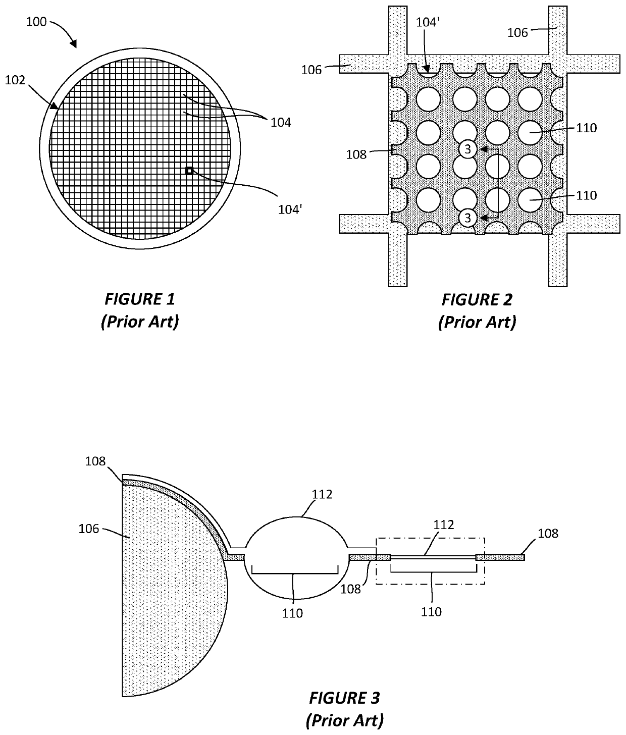
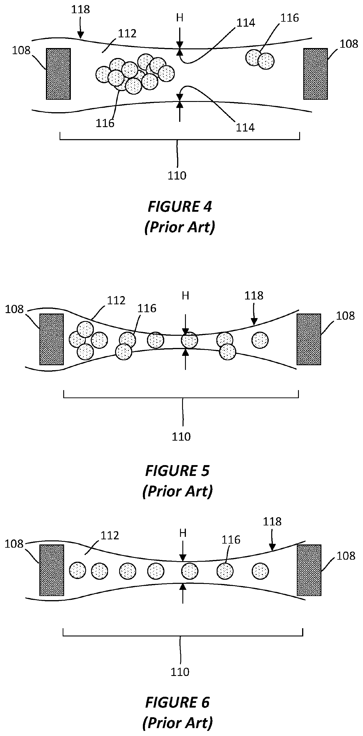
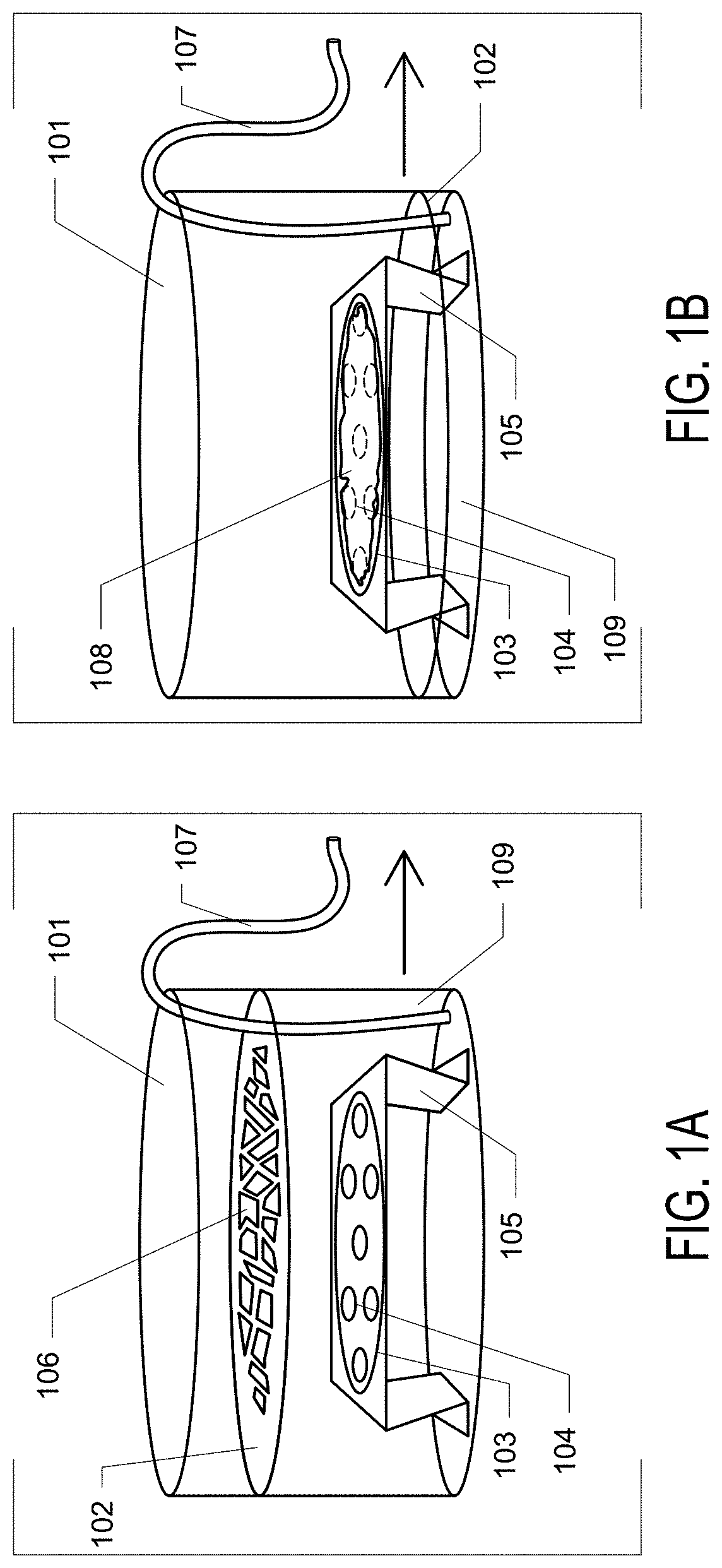
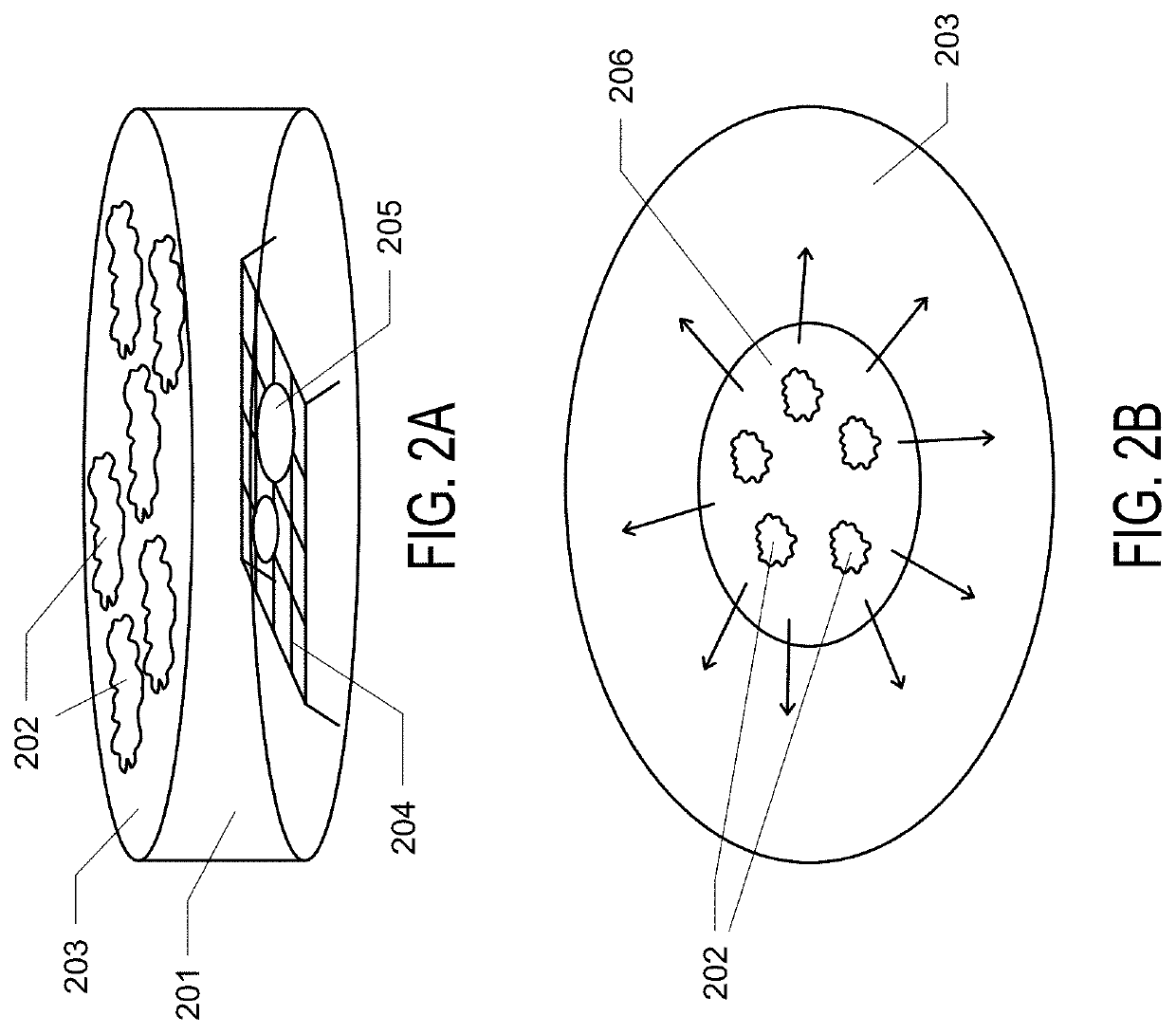
System and method for preparing CRYO-em grids
PatentActiveUS20200363301A1
Innovation
- A method and apparatus that utilize an electromagnetic field (EMF) to re-orient proteins and a system with a sample shaping element and cryogenic vitrifying element to deposit, thin, and vitrify samples on cryo-EM grids, allowing for precise control over sample thickness and orientation, and incorporating a storage device to further randomize protein orientations post-vitrification.
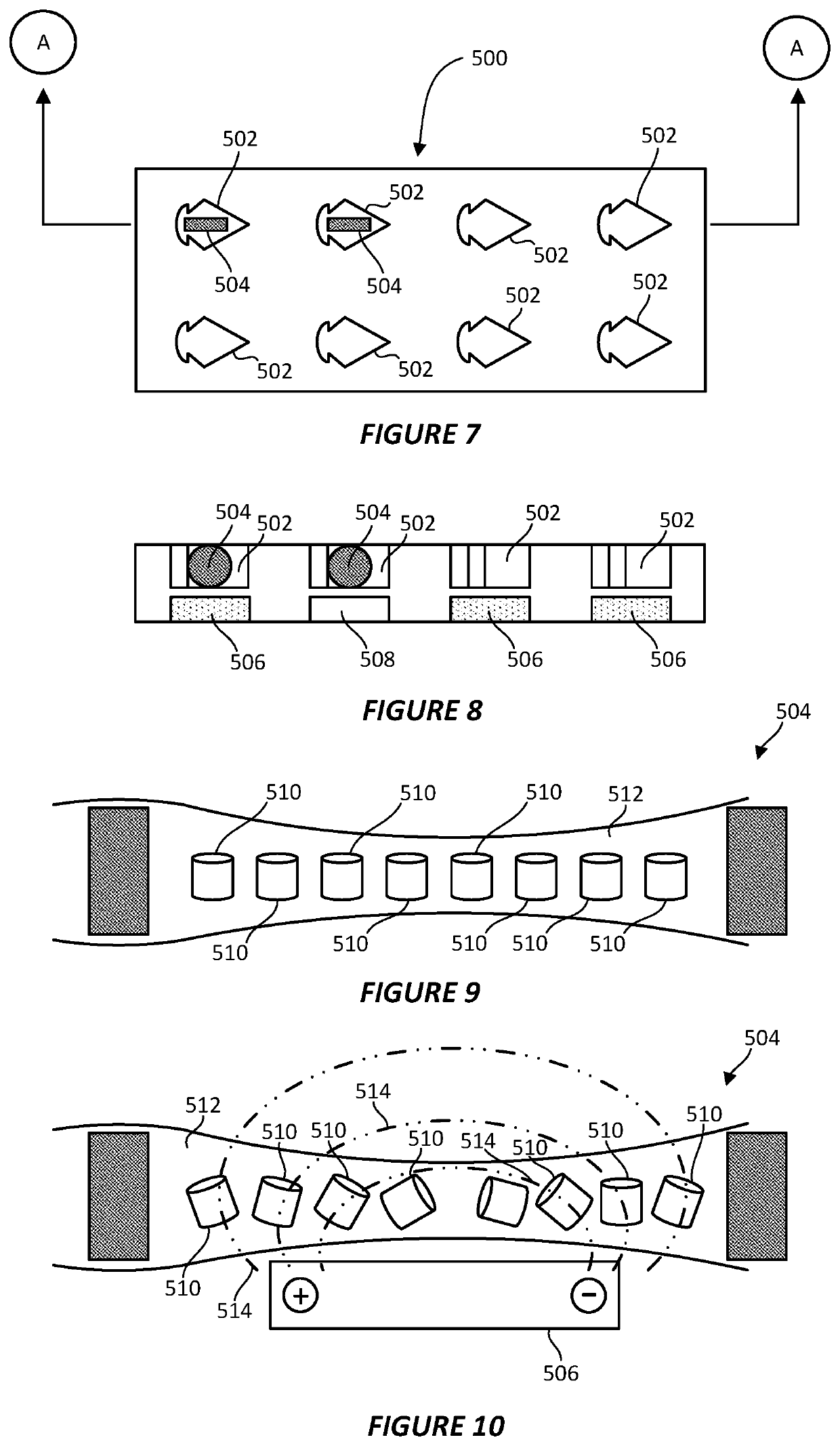
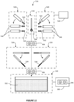
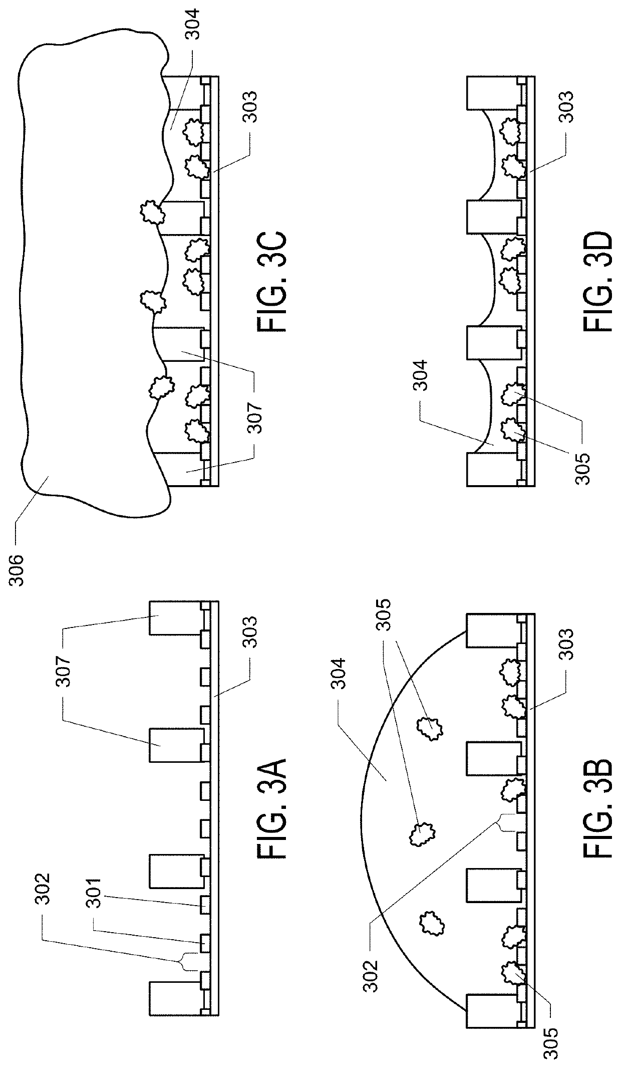
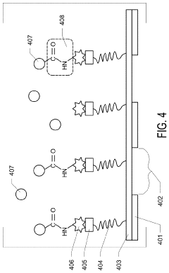
Graphene Oxide Affinity Sample Grids for Cyro-EM
PatentActiveUS20210310910A1
Innovation
- The use of graphene oxide films functionalized with various chemistries for immobilizing diverse target species on cryo-EM grids, combined with polyethylene glycol linkers to position samples optimally away from the air-water interface and substrate, enabling efficient and versatile sample preparation for high-quality cryo-EM imaging.
Sample Preparation Innovations for 2D Materials
Sample preparation represents a critical bottleneck in the application of cryo-electron microscopy (cryo-EM) to 2D material heterostructure interface analysis. Recent innovations have significantly advanced our capabilities in this domain, enabling more precise and reliable structural characterization at the atomic level.
The traditional sample preparation methods for 2D materials often introduce artifacts and contamination that obscure the intricate details of heterostructure interfaces. Novel approaches have emerged to address these limitations, including the development of specialized transfer techniques that minimize surface contamination. The "dry transfer" method, utilizing viscoelastic stamps, has proven particularly effective for creating clean interfaces between different 2D materials without introducing intercalating molecules.
Cryogenic focused ion beam (cryo-FIB) techniques have revolutionized sample preparation by enabling site-specific thinning of heterostructures while maintaining their pristine interfaces. This approach allows researchers to precisely target regions of interest within complex heterostructure stacks, creating electron-transparent windows ideal for high-resolution cryo-EM imaging without compromising the structural integrity of the interfaces.
Another significant advancement involves the use of graphene as an encapsulation layer for 2D material heterostructures. This technique provides dual benefits: it protects sensitive interfaces from oxidation and contamination while serving as an atomically thin support film that minimizes background noise in cryo-EM imaging. The resulting enhancement in signal-to-noise ratio has enabled unprecedented resolution of interface structures.
Plasma cleaning protocols specifically optimized for 2D materials have also emerged as crucial innovations. These protocols effectively remove organic contaminants without damaging the delicate atomic structures of the materials. The precise control of plasma parameters—including gas composition, power, and exposure time—has been refined to accommodate the unique properties of different 2D material combinations.
Automated grid preparation systems have begun to address the reproducibility challenges in cryo-EM sample preparation for 2D materials. These systems provide precise control over vitrification conditions, ensuring consistent ice thickness and minimizing beam-induced damage during imaging. The integration of microfluidic devices with these systems offers promising avenues for high-throughput sample preparation, potentially accelerating the pace of discovery in heterostructure interface research.
Environmental control during transfer and mounting has emerged as another critical innovation area. Advanced transfer chambers that maintain inert atmospheres throughout the sample preparation workflow prevent oxidation and contamination of reactive 2D materials, preserving the native state of heterostructure interfaces for subsequent cryo-EM analysis.
Data Processing Algorithms for Interface Structure Reconstruction
The reconstruction of interface structures from cryo-EM data of 2D material heterostructures presents unique computational challenges that require specialized algorithms. Current data processing approaches leverage both traditional image processing techniques and advanced machine learning methods to extract meaningful structural information from noisy experimental data.
Signal processing algorithms form the foundation of cryo-EM data analysis, with Fourier transforms playing a central role in converting spatial information to frequency domain for more efficient processing. Noise reduction techniques such as Wiener filtering and wavelet transforms have been adapted specifically for the unique noise profiles encountered in cryo-EM imaging of atomically thin interfaces.
Tomographic reconstruction algorithms have evolved significantly to address the specific challenges of 2D material interfaces. Iterative reconstruction methods like SIRT (Simultaneous Iterative Reconstruction Technique) and DART (Discrete Algebraic Reconstruction Technique) have demonstrated superior performance compared to traditional filtered back-projection approaches when dealing with the limited angular sampling often encountered in these specimens.
Machine learning approaches have revolutionized interface structure reconstruction in recent years. Convolutional neural networks trained on simulated data can effectively denoise raw images and enhance contrast at interfaces. Deep learning models such as U-Net architectures have shown remarkable ability to segment different layers in heterostructures and precisely locate interface boundaries with sub-pixel accuracy.
Computational challenges remain significant, particularly for in-situ experiments where real-time processing is desired. GPU acceleration has become standard in processing pipelines, with frameworks like CUDA-enabled TensorFlow providing the computational power needed for complex reconstructions. Distributed computing approaches are increasingly employed for particularly data-intensive analyses.
Validation protocols are essential components of modern reconstruction algorithms. Cross-validation techniques, resolution estimation through Fourier Shell Correlation, and comparison with theoretical models provide confidence metrics for reconstructed structures. These validation approaches are particularly important for heterostructure interfaces where atomic-scale precision is required to understand physical properties.
Recent algorithmic innovations include multi-scale approaches that can simultaneously resolve features at different length scales, particularly valuable for capturing both the extended moiré patterns and local atomic arrangements at interfaces. Phase retrieval algorithms have also been refined to better handle the weak phase contrast typical in cryo-EM imaging of 2D materials.
Unlock deeper insights with PatSnap Eureka Quick Research — get a full tech report to explore trends and direct your research. Try now!
Generate Your Research Report Instantly with AI Agent
Supercharge your innovation with PatSnap Eureka AI Agent Platform!