Cryo-EM For Imaging Liquid Crystal Phases At Nanoscale
AUG 27, 20259 MIN READ
Generate Your Research Report Instantly with AI Agent
Patsnap Eureka helps you evaluate technical feasibility & market potential.
Cryo-EM Nanoscale Imaging Background and Objectives
Cryogenic electron microscopy (Cryo-EM) has emerged as a revolutionary technique in the field of structural biology, enabling researchers to visualize biological macromolecules at near-atomic resolution. Over the past decade, this technology has evolved significantly, transitioning from a specialized tool to a mainstream method for studying complex molecular structures. The application of Cryo-EM to liquid crystal phases represents a particularly promising frontier, as these materials exhibit unique structural properties at the nanoscale that are crucial for various technological applications.
The historical development of Cryo-EM began in the 1980s, but significant breakthroughs occurred in the 2010s with the advent of direct electron detectors and improved image processing algorithms. These advancements dramatically enhanced resolution capabilities, leading to the 2017 Nobel Prize in Chemistry being awarded to the pioneers of this technology. The technique's evolution continues today, with ongoing improvements in sample preparation, detector technology, and computational methods.
Liquid crystals, materials that exhibit properties between those of conventional liquids and solid crystals, have been extensively studied for decades due to their applications in display technologies, sensors, and biomimetic materials. However, understanding their nanoscale organization and phase transitions has remained challenging due to limitations in imaging techniques. Traditional methods such as X-ray diffraction provide averaged structural information but lack the spatial resolution to visualize local defects and domain boundaries that significantly influence material properties.
The primary objective of applying Cryo-EM to liquid crystal phases is to develop a comprehensive understanding of their nanoscale organization, phase transitions, and response to external stimuli. This includes visualizing the formation and dynamics of defects, characterizing domain structures, and elucidating the molecular arrangements that give rise to unique optical and mechanical properties.
Current technological trends indicate a growing convergence between materials science and advanced imaging techniques. The application of Cryo-EM to soft matter systems represents a natural extension of this trend, potentially enabling unprecedented insights into the structure-property relationships of liquid crystalline materials. This approach may reveal previously unobservable features that could inform the design of next-generation materials with tailored properties.
The ultimate goal is to establish Cryo-EM as a routine analytical tool for liquid crystal research, capable of providing real-space structural information with nanometer resolution under conditions that preserve the native state of these sensitive materials. Achieving this would bridge a critical gap in our understanding of soft matter physics and potentially accelerate innovation in fields ranging from flexible electronics to drug delivery systems.
Market Applications for Liquid Crystal Phase Visualization
The market for liquid crystal phase visualization at the nanoscale using Cryo-EM technology spans multiple high-value sectors, with applications continuing to expand as the technology matures. This advanced imaging capability addresses critical needs across scientific research and industrial applications where understanding molecular organization is paramount.
In the pharmaceutical industry, Cryo-EM visualization of liquid crystal phases offers unprecedented insights into drug delivery systems. Lipid-based drug carriers often form liquid crystalline structures that determine release kinetics and bioavailability. Companies like Pfizer and Merck are investing in this technology to optimize formulations for controlled release medications, potentially reducing dosing frequency and improving patient compliance.
The electronics sector represents another significant market, particularly for display technology manufacturers. Understanding liquid crystal phase transitions at the nanoscale directly impacts the development of next-generation displays with improved response times, viewing angles, and energy efficiency. Samsung and LG Display have established research programs utilizing advanced imaging techniques to refine their LCD and OLED technologies.
Cosmetics and personal care products frequently incorporate liquid crystalline structures to enhance stability and delivery of active ingredients. L'Oréal and Estée Lauder employ advanced imaging to develop formulations with improved texture, shelf-life, and efficacy. The ability to visualize how emollients and active ingredients organize at the nanoscale translates to superior product performance.
The food industry benefits from liquid crystal phase visualization in developing structured food systems with controlled texture and release properties. Nestlé and Unilever utilize this knowledge to create products with enhanced mouthfeel, stability, and nutrient bioavailability. The market for functional foods with engineered microstructures continues to grow rapidly.
Materials science represents perhaps the broadest application area, with liquid crystal polymers finding use in high-performance applications from aerospace to medical devices. Companies like DuPont and BASF leverage nanoscale visualization to develop materials with tailored mechanical, thermal, and optical properties.
Academic research institutions constitute a significant market segment, with universities and government laboratories investing in Cryo-EM capabilities to advance fundamental understanding of soft matter physics and self-assembly processes. This research feeds the innovation pipeline for commercial applications across all previously mentioned sectors.
The global market for advanced characterization tools for soft materials, including liquid crystal phases, is projected to grow substantially as industries increasingly recognize the competitive advantage gained through molecular-level understanding of their products and processes.
Technical Challenges in Cryo-EM Liquid Crystal Imaging
Despite significant advancements in cryo-electron microscopy (cryo-EM) techniques, imaging liquid crystal phases at nanoscale presents several formidable technical challenges. The inherent nature of liquid crystals—possessing both fluid-like mobility and crystalline ordering—creates fundamental difficulties for conventional cryo-EM methodologies designed primarily for stable biological specimens.
Sample preparation represents perhaps the most critical challenge. Liquid crystals are extremely sensitive to temperature changes, and the rapid vitrification process required for cryo-EM can disrupt their native phase structure. The cooling rate must be precisely controlled to prevent crystallization of water and formation of ice crystals, which can severely damage the delicate liquid crystal architecture. Additionally, the interaction between liquid crystals and supporting films or grids can induce artificial ordering or phase transitions at interfaces.
Beam-induced damage poses another significant obstacle. Liquid crystal structures are particularly susceptible to radiation damage, with electron doses as low as 1-5 e-/Ų potentially causing structural alterations. This necessitates low-dose imaging techniques that inherently produce noisy data with reduced signal-to-noise ratios, complicating subsequent image processing and interpretation.
The dynamic nature of liquid crystals further complicates imaging efforts. Even in vitrified states, some molecular mobility may persist, leading to blurring effects during image acquisition. This mobility varies across different liquid crystal phases, with nematic phases presenting greater challenges than more ordered smectic or columnar phases due to their higher degree of molecular freedom.
Image interpretation and reconstruction present computational challenges unique to liquid crystal systems. Unlike protein structures with discrete molecular boundaries, liquid crystals exhibit continuous density distributions with subtle variations in molecular orientation. Conventional single-particle analysis algorithms are poorly suited to these systems, requiring specialized computational approaches for accurate structural determination.
Contrast generation represents another technical hurdle. Liquid crystals typically consist of carbon-based molecules with minimal intrinsic contrast in electron microscopy. Phase contrast techniques must be optimized specifically for these materials, often requiring defocus values that balance contrast enhancement against resolution limitations.
Validation of cryo-EM results for liquid crystals remains problematic due to limited complementary techniques at comparable resolution. While X-ray diffraction provides valuable information on periodic structures, it lacks the direct visualization capabilities of cryo-EM, making cross-validation challenging for novel liquid crystal architectures.
Addressing these technical challenges requires interdisciplinary approaches combining advances in sample preparation protocols, detector technology, computational methods, and correlative imaging techniques. Recent developments in direct electron detectors, phase plates, and machine learning-based image processing offer promising pathways toward overcoming these obstacles.
Current Methodologies for Liquid Crystal Nanoscale Imaging
01 High-resolution imaging techniques in Cryo-EM
Advanced techniques have been developed to achieve high-resolution imaging in cryo-electron microscopy. These techniques involve specialized sample preparation, image processing algorithms, and hardware optimizations that collectively enhance the resolution capabilities of cryo-EM systems. Recent innovations have pushed resolution boundaries to near-atomic levels, allowing researchers to visualize molecular structures with unprecedented clarity.- High-resolution imaging techniques in Cryo-EM: Advanced techniques have been developed to achieve high-resolution imaging in Cryo-EM. These techniques involve specialized sample preparation, image processing algorithms, and hardware optimizations that collectively enhance the resolution of structural data. By improving signal-to-noise ratio and reducing beam-induced damage, these methods enable visualization of biological macromolecules at near-atomic resolution, allowing researchers to elucidate detailed molecular structures and mechanisms.
- Sample preparation methods for improved resolution: Innovative sample preparation methods significantly impact Cryo-EM imaging resolution. These include vitrification techniques that preserve specimens in their native state, grid optimization strategies that ensure uniform ice thickness, and specimen purification approaches that enhance homogeneity. Advanced preparation protocols minimize artifacts and contamination while maximizing particle distribution and orientation diversity, which are critical factors for achieving high-resolution reconstructions of biological structures.
- Computational methods for resolution enhancement: Sophisticated computational methods play a crucial role in enhancing Cryo-EM resolution. These include advanced image processing algorithms, machine learning approaches for particle picking and classification, and refined 3D reconstruction techniques. Motion correction, contrast transfer function estimation, and particle alignment algorithms help overcome the inherent limitations of cryo-electron microscopy data. These computational tools enable researchers to extract maximum structural information from noisy, low-contrast images and achieve near-atomic resolution reconstructions.
- Hardware innovations for resolution improvement: Hardware innovations have significantly advanced Cryo-EM imaging resolution capabilities. These include the development of direct electron detectors with improved quantum efficiency, phase plates that enhance contrast without defocusing, and more stable microscope stages that reduce specimen drift. Energy filters and aberration correctors further improve image quality by removing inelastically scattered electrons and correcting lens imperfections. These hardware advancements collectively enable researchers to achieve sub-2 Å resolution for well-behaved samples.
- Integration of Cryo-EM with complementary techniques: Integrating Cryo-EM with complementary structural biology techniques enhances overall resolution and structural insights. Hybrid approaches combining Cryo-EM with X-ray crystallography, NMR spectroscopy, or mass spectrometry provide more comprehensive structural information. Cross-validation between different methods improves confidence in structural models and helps resolve ambiguities. Additionally, correlative light and electron microscopy approaches enable researchers to connect structural information with functional data, providing a more complete understanding of biological systems at multiple resolution scales.
02 Sample preparation methods for improved resolution
Specialized sample preparation methods significantly impact cryo-EM imaging resolution. These methods include vitrification techniques that preserve biological specimens in their native state, grid optimization strategies, and specimen thinning approaches. Proper sample preparation minimizes artifacts, reduces background noise, and enhances contrast, all of which contribute to achieving higher resolution structural data in cryo-EM imaging.Expand Specific Solutions03 Image processing and computational methods
Advanced computational algorithms and image processing techniques play a crucial role in enhancing cryo-EM resolution. These include motion correction, contrast transfer function estimation, particle picking, 3D reconstruction, and refinement methods. Machine learning and artificial intelligence approaches are increasingly being applied to improve signal-to-noise ratios and extract maximum structural information from cryo-EM datasets, pushing resolution limits beyond what was previously achievable.Expand Specific Solutions04 Hardware innovations for resolution enhancement
Hardware advancements have significantly contributed to improving cryo-EM imaging resolution. These innovations include direct electron detectors with improved quantum efficiency, phase plates for enhanced contrast, aberration correctors, and more stable microscope platforms with reduced mechanical and electrical noise. Energy filters and specialized electron optics further enhance signal quality, collectively enabling researchers to achieve sub-2 Ångström resolution in structural studies.Expand Specific Solutions05 Applications of high-resolution cryo-EM in structural biology
High-resolution cryo-EM imaging has revolutionized structural biology by enabling the visualization of complex biological assemblies that were previously challenging to study. These applications include determining the structures of membrane proteins, large macromolecular complexes, viruses, and cellular organelles. The improved resolution capabilities have advanced drug discovery efforts, vaccine development, and fundamental understanding of biological mechanisms at the molecular level.Expand Specific Solutions
Leading Research Groups and Industry Stakeholders
The cryo-electron microscopy (cryo-EM) market for liquid crystal phase imaging at nanoscale is in an early growth stage, with significant research momentum but limited commercial maturity. The global market is expanding as part of the broader electron microscopy sector, valued at approximately $4 billion, with specialized cryo-EM applications representing a growing niche. Leading academic institutions (University of Washington, Max Planck Society, Oxford University) are driving fundamental research, while specialized equipment manufacturers (FEI Co., DENSsolutions, Quantifoil Micro Tools) are developing enabling technologies. The competitive landscape features collaboration between research institutions and commercial entities, with companies like FEI (now part of Thermo Fisher) providing high-end instrumentation while specialized firms like MiTeGen and Ion Dx develop complementary technologies for sample preparation and analysis.
FEI Co.
Technical Solution: FEI Co. (now part of Thermo Fisher Scientific) has developed advanced cryo-electron microscopy (cryo-EM) solutions specifically optimized for liquid crystal phase imaging at nanoscale. Their Titan Krios platform incorporates direct electron detectors with high detective quantum efficiency (DQE) and automated data collection workflows that enable high-resolution imaging of beam-sensitive liquid crystal samples. The company has pioneered low-dose imaging techniques that minimize beam damage while maintaining sufficient contrast to resolve the nanoscale ordering of liquid crystal phases. Their technology integrates sophisticated image processing algorithms that enhance the signal-to-noise ratio in captured images, allowing researchers to visualize the molecular arrangements within various liquid crystal mesophases. FEI's systems also feature specialized sample preparation tools that maintain the native state of liquid crystal structures during the vitrification process, preserving their delicate organizational features for subsequent imaging.
Strengths: Superior resolution capabilities down to sub-nanometer level; integrated workflow from sample preparation to image analysis; advanced beam damage mitigation strategies. Weaknesses: High equipment costs limiting accessibility; requires significant technical expertise to operate effectively; sample preparation remains challenging for certain liquid crystal compositions.
Quantifoil Micro Tools GmbH
Technical Solution: Quantifoil has developed specialized holey carbon film supports specifically designed for cryo-EM imaging of liquid crystal phases. Their R series grids feature precisely engineered hole patterns with controlled spacing and diameter that provide optimal support for liquid crystal samples while minimizing background noise. The company has pioneered hydrophilization treatments that enhance the spreading of liquid crystal samples across the grid surface, ensuring uniform ice thickness and improved imaging quality. Their UltrAuFoil® gold grids offer superior thermal conductivity and mechanical stability compared to traditional carbon supports, reducing beam-induced motion during imaging of sensitive liquid crystal phases. Quantifoil has also developed specialized surface functionalization approaches that help maintain the native organization of liquid crystal samples during the plunge-freezing process, preserving their mesophase structures. Their latest innovations include grids with nanopatterned surfaces that can guide the orientation of liquid crystal domains, facilitating more systematic structural analysis.
Strengths: Exceptional sample support stability reducing motion artifacts; precise control over hole geometry optimized for liquid crystal imaging; gold supports minimize charging effects. Weaknesses: Higher cost compared to standard EM grids; some surface treatments may potentially interact with certain liquid crystal compositions; requires optimization for specific liquid crystal systems.
Key Breakthroughs in Cryo-EM Sample Preparation
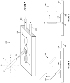
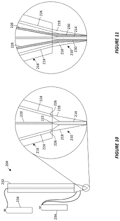
System and method for preparing cryo-em grids
PatentActiveUS20200303162A1
Innovation
- A cryogenically-cooled sample preparation apparatus with a movable and rotatable sample dispenser that automatically deposits and vitrifies liquid samples on a grid, eliminating the need for manual handling and blotting, and allowing for precise control of ice layer thickness through adjustable deposition rates and cryogen circulation.
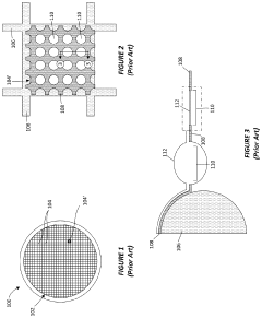
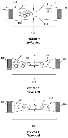
Thin-ice grid assembly for cryo-electron microscopy
PatentInactiveUS20160351374A1
Innovation
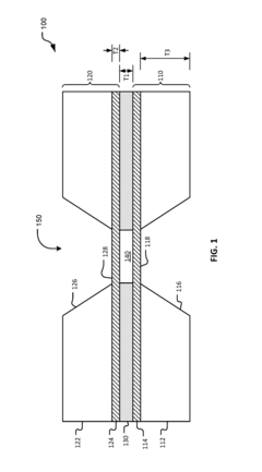
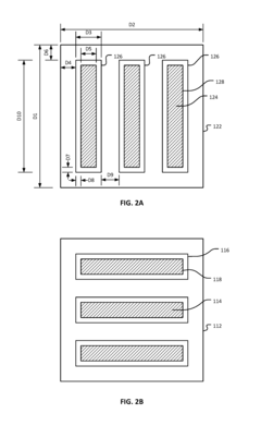
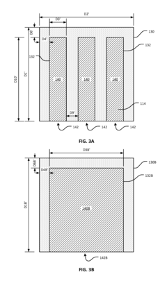
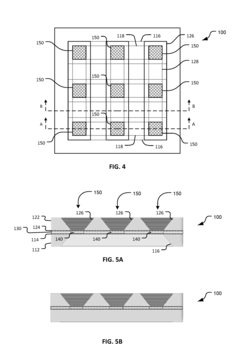
- A grid assembly for cryo-EM is developed, comprising two support members with electron-transparent layers and a rigid spacer layer, allowing precise control of ice thickness between them, enabling consistent vitrification and efficient imaging.
Materials Science Integration with Cryo-EM Techniques
The integration of materials science with cryo-electron microscopy (cryo-EM) represents a significant advancement in nanoscale imaging capabilities, particularly for studying liquid crystal phases. This convergence has enabled researchers to overcome traditional limitations in visualizing dynamic molecular arrangements that define liquid crystalline states.
Materials science principles provide the theoretical framework for understanding the complex phase behaviors of liquid crystals, while cryo-EM offers the technical means to capture these structures in their native state. The rapid freezing process inherent to cryo-EM preserves the delicate molecular ordering of liquid crystals without introducing crystallization artifacts that would otherwise disrupt their natural arrangement.
Recent developments have focused on optimizing sample preparation protocols specifically for liquid crystalline materials. These include controlled vitrification techniques that maintain the orientational order of mesophases while preventing ice crystal formation. Additionally, specialized grid treatments have been developed to manage the unique surface interactions between liquid crystals and support films, ensuring minimal distortion during the freezing process.
The integration has also driven innovations in image processing algorithms tailored to the particular challenges of liquid crystal data. These computational approaches can distinguish between different mesophases and quantify orientational parameters with unprecedented precision, extracting structural information that was previously inaccessible through conventional imaging methods.
Cross-disciplinary collaboration between materials scientists and structural biologists has accelerated method development, with techniques originally designed for biological macromolecules now being adapted for soft matter physics applications. This knowledge transfer has been particularly valuable in developing low-dose imaging protocols that minimize beam-induced damage to radiation-sensitive liquid crystal samples.
Temperature-controlled sample holders represent another important technical advancement, allowing researchers to study phase transitions in situ. These specialized devices maintain precise thermal conditions during imaging, enabling the observation of temperature-dependent structural changes in liquid crystal phases at nanometer resolution.
The materials science perspective has also informed the interpretation of cryo-EM data, providing theoretical models that help explain the observed molecular arrangements in terms of fundamental physical principles. This interpretative framework enhances the value of imaging data by connecting visual observations to underlying thermodynamic and kinetic processes governing liquid crystal behavior.
Data Processing Algorithms for Liquid Crystal Structure Analysis
Data processing algorithms play a crucial role in extracting meaningful structural information from cryo-EM images of liquid crystal phases at the nanoscale. The complexity of liquid crystal structures, characterized by their orientational order and positional disorder, requires sophisticated computational approaches to accurately interpret the acquired data.
Traditional image processing techniques often fall short when applied to liquid crystal samples due to the inherent molecular mobility and varying degrees of order present in different mesophases. Recent advancements have led to the development of specialized algorithms that can account for these unique characteristics, enabling more precise structural analysis.
Fourier transform-based methods represent the foundation of many liquid crystal structure analysis algorithms. These techniques transform spatial domain information into frequency domain representations, allowing researchers to identify periodic patterns and orientational order parameters. Enhanced variants incorporating wavelet transforms have shown particular promise in detecting local structural variations within heterogeneous liquid crystal samples.
Machine learning approaches have revolutionized data processing for cryo-EM liquid crystal imaging. Convolutional neural networks (CNNs) trained on simulated liquid crystal structures can now automatically classify different mesophases and extract order parameters with minimal human intervention. These algorithms have demonstrated superior performance in distinguishing subtle structural differences between similar liquid crystal phases.
Noise reduction algorithms specifically optimized for cryo-EM liquid crystal data have emerged as essential preprocessing tools. These algorithms can differentiate between thermal fluctuations inherent to liquid crystals and instrumental noise, preserving critical structural information while enhancing image clarity. Adaptive filtering techniques that consider local structural contexts have proven particularly effective.
Tomographic reconstruction algorithms adapted for liquid crystal samples enable three-dimensional visualization of complex mesophases. These methods account for the anisotropic nature of liquid crystals by incorporating orientation-dependent scattering models into the reconstruction process. Recent implementations utilizing GPU acceleration have significantly reduced processing times, enabling near real-time structural analysis.
Integration of molecular dynamics simulations with experimental data processing has created powerful hybrid algorithms that can validate structural models against cryo-EM observations. These approaches iteratively refine molecular arrangements until simulated electron density maps converge with experimental data, providing insights into dynamic behaviors not directly observable through imaging alone.
Unlock deeper insights with Patsnap Eureka Quick Research — get a full tech report to explore trends and direct your research. Try now!
Generate Your Research Report Instantly with AI Agent
Supercharge your innovation with Patsnap Eureka AI Agent Platform!
