Cryo-EM For Imaging Energy Materials Under Controlled Atmospheres
AUG 27, 202510 MIN READ
Generate Your Research Report Instantly with AI Agent
Patsnap Eureka helps you evaluate technical feasibility & market potential.
Cryo-EM Technology Evolution and Research Objectives
Cryogenic electron microscopy (Cryo-EM) has undergone remarkable evolution since its inception in the 1980s, transforming from a niche technique primarily used in structural biology to a versatile imaging method with expanding applications across multiple scientific domains. The fundamental breakthrough came with the development of vitrification methods that allowed biological samples to be preserved in their native state without crystallization damage. This innovation laid the groundwork for subsequent technological advancements in both hardware and software components.
The past decade has witnessed unprecedented progress in Cryo-EM technology, marked by significant improvements in electron detectors, particularly direct electron detectors that dramatically enhance signal-to-noise ratios. Concurrent advancements in image processing algorithms and computational methods have enabled researchers to achieve near-atomic resolution imaging, revolutionizing our understanding of complex molecular structures.
The application of Cryo-EM to energy materials represents a relatively recent but rapidly growing research direction. Traditional characterization techniques often fail to capture the dynamic processes and structural changes occurring in energy materials under operating conditions. Cryo-EM offers unique capabilities to observe these materials at nanoscale resolution while maintaining controlled atmospheric conditions, providing insights into degradation mechanisms, interface phenomena, and reaction pathways.
Current research objectives in Cryo-EM for energy materials focus on several key areas. First, developing specialized sample preparation techniques that preserve the native state of energy materials while allowing for controlled atmospheric conditions during imaging. Second, enhancing resolution capabilities to visualize atomic-level structural changes during energy conversion and storage processes. Third, integrating in-situ capabilities to observe dynamic processes in real-time under various environmental conditions.
The field aims to bridge fundamental understanding with practical applications by correlating structural characteristics with functional properties of energy materials. This includes investigating battery electrode materials during charge-discharge cycles, catalyst surfaces during reaction processes, and membrane materials for fuel cells and electrolyzers under operating conditions.
Technical challenges being addressed include minimizing beam damage to sensitive energy materials, developing specialized holders and chambers for controlled atmosphere experiments, and creating analytical frameworks to extract quantitative information from the acquired images. The ultimate goal is to establish Cryo-EM as an indispensable characterization tool that provides unique insights into energy materials behavior, thereby accelerating the development of next-generation energy technologies with improved efficiency, durability, and performance.
Market Applications for Atmospheric-Controlled Energy Materials Imaging
The atmospheric-controlled cryo-EM imaging technology for energy materials addresses critical market needs across multiple sectors, with particularly strong applications in battery development and fuel cell optimization. The global lithium-ion battery market, currently experiencing rapid growth, requires advanced characterization techniques to overcome performance limitations and safety concerns. Atmospheric-controlled cryo-EM enables researchers to observe battery materials in their native environments, providing crucial insights into degradation mechanisms and interface dynamics that traditional imaging methods cannot capture.
In the electric vehicle sector, manufacturers are seeking longer-lasting, faster-charging batteries with higher energy densities. This imaging technology allows for direct visualization of solid-electrolyte interphase formation and lithium dendrite growth under controlled conditions, addressing key challenges in next-generation battery development. The technology's ability to preserve sensitive samples in their reactive states while maintaining atmospheric control represents a significant advancement for industrial R&D departments working on improving battery cycle life and safety.
Hydrogen fuel cell development represents another substantial market application, where catalyst degradation and membrane electrode assembly performance can be studied at unprecedented resolution. The technology enables visualization of platinum nanoparticle catalysts under reactive gas environments, providing critical information for reducing platinum loading and improving catalyst utilization - addressing a major cost barrier in fuel cell commercialization.
Solar cell manufacturing can also benefit significantly from atmospheric-controlled cryo-EM, particularly for perovskite solar cells where moisture sensitivity has been a persistent challenge. The ability to image these materials under controlled humidity conditions provides manufacturers with insights to develop more stable encapsulation methods and moisture-resistant formulations, potentially extending product lifetimes.
Energy storage grid applications represent an emerging market segment where understanding degradation mechanisms in flow batteries and grid-scale storage solutions is essential for improving reliability. The technology's capability to image materials under electrochemical cycling conditions while maintaining atmospheric control provides valuable data for extending operational lifetimes of these critical infrastructure components.
Carbon capture technologies and next-generation nuclear materials also represent growing application areas, where material-gas interactions must be thoroughly understood for safety and efficiency. The ability to directly visualize how these materials interact with specific gas environments at near-atomic resolution provides crucial data for optimizing designs and improving performance in these emerging energy sectors.
Current Challenges in Cryo-EM for Energy Materials Characterization
Despite significant advancements in cryo-electron microscopy (cryo-EM) for energy materials characterization, several critical challenges persist that limit its full potential in this domain. The primary obstacle remains sample preparation under controlled atmospheres, as many energy materials are highly reactive with air and moisture. Traditional cryo-EM sample preparation techniques expose specimens to ambient conditions, potentially altering their native structures and chemical compositions before imaging can occur.
Resolution limitations continue to challenge researchers when examining energy materials at atomic scales. While recent technological improvements have pushed resolution boundaries, achieving consistent sub-angstrom resolution for complex heterogeneous energy materials remains difficult. This is particularly problematic when investigating interfaces and defects that often determine critical performance characteristics in batteries, fuel cells, and catalysts.
Beam damage presents another significant hurdle, as electron beam interactions can induce structural changes and chemical reactions in sensitive energy materials. This is especially problematic for organic components in hybrid energy materials and lithium-containing compounds, where even low electron doses can trigger substantial alterations to the specimen during observation.
The dynamic nature of energy materials poses unique challenges for cryo-EM imaging. Many energy conversion and storage processes involve structural and chemical changes that occur rapidly and continuously. Capturing these transformations in real-time while maintaining cryogenic conditions and controlled atmospheres requires sophisticated in-situ capabilities that are not yet fully developed.
Data interpretation complexities arise from the heterogeneity of energy materials. Distinguishing between intrinsic material features and artifacts introduced during sample preparation or imaging remains challenging. Advanced computational methods for image processing and 3D reconstruction are needed to extract meaningful information from noisy and complex datasets.
Technical integration issues exist between cryo-EM and complementary characterization techniques. Correlative approaches combining cryo-EM with spectroscopic methods would provide comprehensive structural and chemical information, but practical implementation faces significant engineering challenges related to maintaining sample integrity across different instruments.
Cost and accessibility barriers limit widespread adoption of advanced cryo-EM techniques in energy materials research. High-end instruments with specialized capabilities for controlled atmosphere imaging remain prohibitively expensive for many research institutions, creating disparities in research capabilities across the field.
Existing Methodologies for Controlled-Atmosphere Cryo-EM
01 Sample preparation techniques for Cryo-EM
Various methods have been developed for preparing biological samples for cryogenic electron microscopy imaging. These techniques focus on preserving the native structure of specimens during the freezing process, including vitrification methods that prevent ice crystal formation. Advanced sample holders and grid designs enable better specimen stability during imaging, while specialized preparation protocols have been developed for different types of biological materials such as proteins, viruses, and cellular structures.- Sample preparation techniques for Cryo-EM: Various methods and devices for preparing biological samples for cryogenic electron microscopy imaging. These techniques focus on preserving the native structure of biological specimens through rapid freezing processes that prevent ice crystal formation. The preparation methods include vitrification, grid preparation, and specialized sample holders that maintain ultra-low temperatures during imaging. These techniques are crucial for achieving high-resolution structural information of biomolecules in their near-native states.
- Image processing and reconstruction algorithms: Advanced computational methods for processing and reconstructing three-dimensional structures from Cryo-EM data. These algorithms address challenges such as low signal-to-noise ratio, beam-induced motion, and heterogeneity in sample conformations. Machine learning and artificial intelligence approaches are increasingly being applied to enhance image quality, automate particle picking, and improve resolution in the final reconstructions. These computational advances have been instrumental in the recent revolution in structural biology using Cryo-EM.
- Hardware innovations for Cryo-EM microscopes: Technical improvements in electron microscope hardware specifically designed for cryogenic applications. These innovations include advanced electron detectors with improved sensitivity and speed, more stable cold stages that reduce specimen drift, and enhanced electron optics that minimize aberrations. Other hardware developments focus on automation systems for high-throughput data collection and specialized components that maintain the vitrified state of biological samples during imaging sessions.
- Applications in structural biology and drug discovery: Implementation of Cryo-EM techniques for determining the structures of biological macromolecules and their complexes to facilitate drug discovery and development. This approach allows visualization of protein-drug interactions at near-atomic resolution, enabling structure-based drug design. Cryo-EM has become particularly valuable for studying membrane proteins, large molecular assemblies, and conformationally heterogeneous samples that are challenging to analyze by traditional structural methods like X-ray crystallography.
- Integration with complementary techniques: Methods for combining Cryo-EM with other analytical techniques to enhance structural information and functional understanding of biological systems. These hybrid approaches include correlative light and electron microscopy, integration with mass spectrometry, and computational modeling. By merging data from multiple experimental methods, researchers can obtain more comprehensive insights into molecular structures and dynamics across different spatial and temporal scales, overcoming the limitations of any single technique.
02 Image processing and reconstruction algorithms
Sophisticated computational methods are essential for processing Cryo-EM data and reconstructing three-dimensional structures from two-dimensional images. These algorithms include motion correction to account for beam-induced movement, contrast transfer function estimation, particle picking, classification, and 3D reconstruction techniques. Machine learning and artificial intelligence approaches have been integrated to enhance image quality, improve signal-to-noise ratios, and automate parts of the reconstruction workflow.Expand Specific Solutions03 Hardware innovations for Cryo-EM systems
Technical advancements in Cryo-EM hardware have significantly improved imaging capabilities. These innovations include direct electron detectors with higher sensitivity and faster frame rates, improved electron sources for better coherence and brightness, more stable specimen stages to reduce drift, and automated data collection systems. Energy filters and phase plates have been developed to enhance contrast in biological specimens, while improvements in vacuum systems and temperature control maintain sample integrity during imaging.Expand Specific Solutions04 Integration with other analytical techniques
Cryo-EM is increasingly being combined with complementary analytical methods to provide more comprehensive structural information. These hybrid approaches include correlative light and electron microscopy (CLEM), which combines fluorescence microscopy with Cryo-EM, integration with mass spectrometry for molecular identification, and combination with X-ray crystallography or NMR spectroscopy for multi-scale structural analysis. These integrated approaches enable researchers to connect structural information with functional insights across different resolution scales.Expand Specific Solutions05 Time-resolved and dynamic Cryo-EM methods
Emerging techniques allow for the capture of biological processes in action rather than just static structures. These approaches include time-resolved Cryo-EM methods that trap molecules at different functional states, microfluidic devices for rapid mixing and freezing of samples at defined time points, and computational approaches for sorting heterogeneous conformational states. These innovations enable researchers to visualize dynamic molecular processes and conformational changes that are critical for understanding biological function.Expand Specific Solutions
Leading Research Institutions and Equipment Manufacturers
The cryo-electron microscopy (cryo-EM) market for energy materials imaging under controlled atmospheres is currently in its growth phase, with an expanding market size driven by increasing demand for advanced material characterization techniques. The technology is approaching maturity but still evolving, with key players including established microscopy companies like FEI Co. (now part of Thermo Fisher) and Gatan, Inc., who provide core imaging systems. Specialized component manufacturers such as Protochips, MiTeGen, Quantifoil Micro Tools, and Simple Origin are developing crucial accessories for controlled-environment imaging. Academic institutions (Max Planck Society, Oxford, Harvard, Tsinghua) and research organizations (EMBL, IBS) are advancing fundamental techniques, while commercial applications are being explored by companies like Genentech. The ecosystem demonstrates a collaborative approach between hardware providers, research institutions, and end-users to overcome technical challenges in imaging energy materials under realistic conditions.
FEI Co.
Technical Solution: FEI (now part of Thermo Fisher Scientific) has developed comprehensive cryo-EM solutions for imaging energy materials under controlled atmospheres. Their Titan and Talos microscope platforms incorporate environmental cells that maintain stable gas environments while enabling atomic-resolution imaging. FEI's Direct Electron Detector technology provides exceptional sensitivity for low-dose imaging of beam-sensitive energy materials like lithium-ion battery components and fuel cell catalysts. Their Auto-Loader system enables rapid sample exchange while maintaining cryogenic conditions, critical for studying materials in their native state. FEI's tomography capabilities allow 3D reconstruction of energy material structures under various atmospheric conditions, providing insights into porosity, connectivity, and interface properties. The company's integrated workflow solutions encompass sample preparation, data acquisition, and analysis specifically optimized for energy materials research, enabling correlative studies across multiple length scales.
Strengths: Comprehensive integrated solutions from sample preparation to analysis; industry-leading resolution capabilities; extensive application support network. Weaknesses: High capital investment requirements; complex systems require specialized training; environmental cells may have limitations in extreme conditions.
Protochips, Inc.
Technical Solution: Protochips has pioneered specialized in-situ holders and environmental cells specifically designed for cryo-EM imaging of energy materials under controlled atmospheres. Their Atmosphere system enables precise gas environment control (up to 1 atm pressure) while maintaining high-resolution imaging capabilities. The company's Fusion heating technology allows temperature control from cryogenic to 1200°C, critical for studying phase transitions in battery materials and catalysts. Protochips' Poseidon liquid-cell system permits direct observation of electrochemical processes in liquid electrolytes, enabling visualization of electrode-electrolyte interfaces during operation. Their integrated software platform provides synchronized control of environmental parameters with image acquisition, allowing correlation between material structure changes and environmental conditions. The company's MEMS-based sample supports minimize thermal drift and mechanical vibration, enhancing image stability during environmental transitions.
Strengths: Precise environmental parameter control; specialized solutions for energy material applications; compatibility with multiple microscope platforms. Weaknesses: Limited field of view in some environmental cell configurations; potential for electron beam interactions with gas environments; higher complexity in experimental setup compared to conventional cryo-EM.
Critical Innovations in Sample Preparation and Data Processing
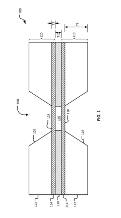
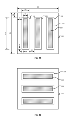
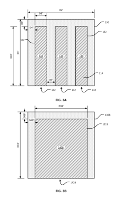
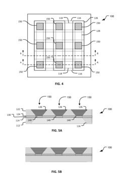
Thin-ice grid assembly for CRYO-electron microscopy
PatentWO2015134575A1
Innovation
- A thin-ice grid assembly is developed, comprising two electron-transparent support members with a rigid spacer layer, allowing precise control of ice thickness between them, enabling consistent vitrification and improved imaging conditions.
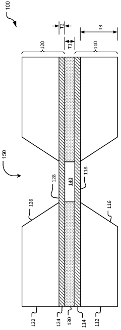
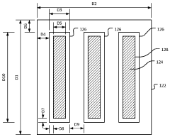
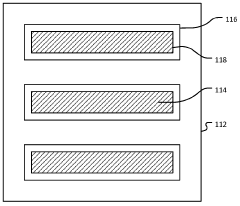
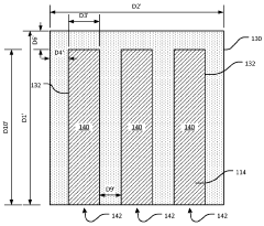
Thin-ice grid assembly for cryo-electron microscopy
PatentInactiveUS20160351374A1
Innovation
- A grid assembly for cryo-EM is developed, comprising two support members with electron-transparent layers and a rigid spacer layer, allowing precise control of ice thickness between them, enabling consistent vitrification and efficient imaging.
Environmental Impact and Sustainability Considerations
The implementation of Cryo-EM for imaging energy materials under controlled atmospheres carries significant environmental implications that must be carefully considered. The technology's environmental footprint primarily stems from its substantial energy consumption requirements, particularly for maintaining ultra-low temperatures necessary for cryogenic conditions. These systems typically operate at temperatures below -150°C, demanding continuous cooling through liquid nitrogen or helium, both of which require energy-intensive production processes.
Laboratory facilities housing Cryo-EM equipment must maintain precise environmental controls, further increasing energy demands. The carbon footprint associated with these operations becomes particularly relevant when considering the technology's scaling for industrial applications in energy materials research. Ironically, while the technology aims to advance sustainable energy solutions, its own energy consumption profile presents a sustainability challenge that requires optimization.
Chemical waste management represents another critical environmental consideration. Sample preparation for Cryo-EM often involves various chemicals, including heavy metals for staining and contrast enhancement, organic solvents, and cryoprotectants. Proper disposal protocols must be established to prevent environmental contamination, particularly as the technology sees wider adoption across research institutions and industrial settings.
The controlled atmospheres essential for accurate material characterization introduce additional environmental considerations. Specialized gases used in these environments may include rare or manufactured gases with varying environmental impacts in their production, transportation, and potential leakage scenarios. Recycling systems for these gases can significantly reduce both environmental impact and operational costs.
From a sustainability perspective, Cryo-EM technology offers promising contributions to green technology development. By enabling precise characterization of energy materials at atomic scales under realistic operating conditions, it accelerates the development of more efficient batteries, fuel cells, and catalysts. These advancements directly support renewable energy adoption and reduced fossil fuel dependence. The environmental benefits of these innovations may ultimately outweigh the environmental costs of the technology itself.
Life cycle assessment (LCA) methodologies should be applied to Cryo-EM systems to quantify their complete environmental impact from manufacturing through operation to eventual decommissioning. This analysis would provide valuable insights for developing more sustainable next-generation instruments with reduced environmental footprints, potentially incorporating energy recovery systems, more efficient cooling technologies, and environmentally friendly sample preparation protocols.
Integration with Complementary Characterization Techniques
The integration of cryo-electron microscopy (cryo-EM) with complementary characterization techniques represents a significant advancement in the comprehensive analysis of energy materials under controlled atmospheres. This synergistic approach enables researchers to obtain multidimensional insights that a single technique cannot provide, thereby enhancing the overall understanding of material properties and behaviors.
X-ray diffraction (XRD) techniques serve as powerful companions to cryo-EM, offering crystallographic information that complements the high-resolution structural data from electron microscopy. When combined, these techniques provide a more complete picture of both crystalline and amorphous phases in energy materials, particularly important for battery electrodes and catalysts where phase transitions occur under different atmospheric conditions.
Spectroscopic methods such as X-ray photoelectron spectroscopy (XPS) and Raman spectroscopy can be effectively integrated with cryo-EM workflows to correlate structural observations with chemical state information. This integration is particularly valuable when studying surface reactions in fuel cells or electrocatalysts, where both structural changes and chemical transformations occur simultaneously under controlled gas environments.
Advanced synchrotron-based techniques, including X-ray absorption spectroscopy (XAS) and X-ray tomography, offer complementary bulk analysis capabilities that extend the primarily surface-sensitive information obtained from cryo-EM. Researchers have developed specialized sample holders and transfer systems that maintain controlled atmospheres while moving specimens between different characterization platforms, preserving the material state across multiple analytical techniques.
In situ and operando measurements represent the frontier of integrated characterization approaches. Recent developments have focused on creating compatible sample environments that allow for sequential or even simultaneous measurements using multiple techniques while maintaining identical atmospheric conditions. For example, specialized cells have been designed to accommodate both electron and X-ray beams, enabling direct correlation between structural evolution observed in cryo-EM and electrochemical performance measured through other techniques.
Computational methods play a crucial role in integrating data from multiple characterization techniques. Machine learning algorithms and multivariate statistical approaches have been developed to correlate information across different length scales and measurement modalities. These computational frameworks help researchers extract meaningful relationships between atomic-scale structures observed in cryo-EM and macroscopic properties measured through complementary techniques.
The integration of cryo-EM with thermal analysis techniques such as differential scanning calorimetry (DSC) and thermogravimetric analysis (TGA) provides valuable insights into the thermodynamic and kinetic aspects of material transformations under controlled atmospheres. This combined approach is particularly important for understanding degradation mechanisms in energy storage materials and the activation processes in heterogeneous catalysts.
Unlock deeper insights with Patsnap Eureka Quick Research — get a full tech report to explore trends and direct your research. Try now!
Generate Your Research Report Instantly with AI Agent
Supercharge your innovation with Patsnap Eureka AI Agent Platform!