Cryo-EM For Imaging Liquid-Containing Nanomaterials
AUG 27, 20259 MIN READ
Generate Your Research Report Instantly with AI Agent
Patsnap Eureka helps you evaluate technical feasibility & market potential.
Cryo-EM Technology Background and Objectives
Cryo-electron microscopy (Cryo-EM) has revolutionized the field of structural biology since its inception in the 1980s. Initially developed for visualizing biological macromolecules, this technique has evolved significantly over the past four decades, with major breakthroughs occurring in the 2010s that led to the Nobel Prize in Chemistry in 2017. The fundamental principle involves flash-freezing samples in vitreous ice, preserving their native structure while allowing electron beam penetration for high-resolution imaging.
The application of Cryo-EM to liquid-containing nanomaterials represents a relatively recent but rapidly growing frontier. Traditional electron microscopy techniques require samples to be in vacuum conditions, making the imaging of liquid phases exceptionally challenging. The development of specialized sample preparation methods and environmental chambers has gradually enabled researchers to overcome these limitations, opening new possibilities for studying nanomaterials in their native liquid environments.
The technological evolution of Cryo-EM has been marked by several key advancements, including improved electron detectors, automated data collection systems, and sophisticated image processing algorithms. These developments have collectively pushed resolution capabilities from nanometer to near-atomic levels, making it possible to visualize intricate structural details of nanomaterials in liquid phases that were previously inaccessible.
Current research trends are focused on enhancing temporal resolution to capture dynamic processes, improving sample preparation techniques to minimize artifacts, and developing correlative approaches that combine Cryo-EM with other analytical methods. The integration of artificial intelligence and machine learning algorithms for image processing and reconstruction represents another significant trend that is accelerating progress in this field.
The primary objectives of Cryo-EM for liquid-containing nanomaterials include: characterizing the structure-function relationships of nanomaterials in liquid environments; understanding dynamic processes such as self-assembly, nucleation, and growth; investigating interfaces between nanomaterials and surrounding liquids; and developing in situ experimental capabilities to observe reactions and transformations in real-time.
Looking forward, the field aims to achieve true atomic resolution imaging of liquid-nanomaterial systems, develop methods for time-resolved studies at microsecond to millisecond timescales, and establish standardized protocols for reliable and reproducible sample preparation. These advancements would significantly impact various fields including materials science, nanotechnology, catalysis, energy storage, and environmental remediation by providing unprecedented insights into nanomaterial behavior in realistic conditions.
Market Applications for Liquid-Containing Nanomaterial Imaging
The market for liquid-containing nanomaterial imaging technologies, particularly those utilizing Cryo-EM, is experiencing significant growth across multiple sectors. Pharmaceutical and biotechnology industries represent the largest market segment, with an estimated market value exceeding $3 billion globally. These industries leverage Cryo-EM imaging for drug discovery processes, particularly in structure-based drug design where visualizing protein-ligand interactions in their native hydrated state provides crucial insights for developing targeted therapeutics.
Materials science and nanotechnology sectors constitute another substantial market, where Cryo-EM enables researchers to visualize liquid-solid interfaces and dynamic processes within nanomaterials. This capability is particularly valuable for developing advanced materials with controlled properties, such as specialized coatings, catalysts, and energy storage materials. The ability to observe nanomaterials in their hydrated state provides unprecedented insights into their behavior under real-world conditions.
The semiconductor industry has recently emerged as a rapidly growing application area. As device dimensions continue to shrink, understanding the behavior of liquid precursors and solutions used in fabrication processes becomes increasingly critical. Cryo-EM offers unique capabilities for imaging these liquid-phase processes at nanoscale resolution, helping manufacturers optimize their production techniques and improve yield rates.
Environmental monitoring and remediation represent an expanding market segment where Cryo-EM imaging of liquid-containing nanomaterials aids in developing more effective filtration systems, pollution detection methods, and water treatment technologies. The ability to visualize contaminants and remediation nanomaterials in aqueous environments provides valuable data for improving environmental protection strategies.
Academic and government research institutions form a stable market base, driving fundamental research that ultimately feeds innovation in commercial applications. These institutions typically focus on advancing the technology itself while exploring novel applications across disciplines including chemistry, biology, and materials science.
Geographically, North America currently dominates the market with approximately 40% share, followed by Europe and Asia-Pacific regions. However, the Asia-Pacific market is projected to grow at the fastest rate over the next five years, driven by increasing R&D investments in China, Japan, and South Korea, particularly in semiconductor and materials science applications.
Market growth is further accelerated by the convergence of Cryo-EM with complementary technologies such as artificial intelligence for image processing and analysis, creating more accessible and powerful analytical tools for researchers and industry professionals working with liquid-containing nanomaterials.
Current Challenges in Cryo-EM Liquid Nanomaterial Analysis
Despite significant advancements in cryo-electron microscopy (cryo-EM) techniques, imaging liquid-containing nanomaterials presents several persistent challenges that hinder comprehensive analysis. The primary obstacle remains sample preparation, as the conventional vitrification process can disrupt the native state of liquid-containing nanomaterials. The rapid freezing required to achieve vitreous ice often causes structural alterations, particularly in systems with complex liquid-solid interfaces or those containing volatile components.
Beam-induced damage represents another critical limitation. The electron beam interaction with liquid components frequently leads to radiolysis, bubble formation, and molecular degradation, compromising the structural integrity of the specimen during imaging. This damage occurs even at cryogenic temperatures and with low-dose imaging protocols, making it particularly problematic for extended data collection sessions necessary for high-resolution reconstructions.
Resolution barriers continue to impede progress in this field. While cryo-EM has achieved near-atomic resolution for many biological macromolecules, liquid-containing nanomaterials often yield lower resolution data due to inherent sample heterogeneity and motion of liquid components. The dynamic nature of liquids, even when vitrified, introduces blurring effects that limit achievable resolution.
Contrast generation presents unique difficulties when imaging liquid-containing nanomaterials. The similar electron densities between vitrified liquid components and surrounding ice matrix often result in poor contrast, making particle detection and alignment challenging. Phase plates and other contrast enhancement technologies have shown promise but remain technically demanding to implement consistently.
Data processing algorithms face significant hurdles when dealing with liquid-containing samples. Current classification and averaging approaches were primarily developed for discrete, rigid particles and struggle with the conformational heterogeneity inherent in liquid-containing systems. The continuous nature of liquid-solid interfaces defies traditional particle-picking strategies and requires specialized computational approaches.
Environmental control during sample transfer and imaging represents another technical challenge. Maintaining the native state of liquid components throughout the workflow demands sophisticated cryo-transfer systems and rigorous temperature management to prevent devitrification or ice contamination. Even minor temperature fluctuations can induce phase transitions that irreversibly alter sample morphology.
Correlative imaging approaches, combining cryo-EM with other analytical techniques, remain underdeveloped for liquid-containing nanomaterials. Integrating spectroscopic or diffraction data with structural information would provide valuable insights but requires complex instrumentation and sample preparation protocols that are not yet standardized.
Current Methodologies for Liquid-Containing Nanomaterial Imaging
01 Sample preparation techniques for Cryo-EM
Various methods for preparing biological samples for cryogenic electron microscopy imaging to preserve their native structure. These techniques include vitrification processes, grid preparation methods, and specialized sample holders that maintain ultra-low temperatures. Proper sample preparation is crucial for achieving high-resolution structural information while minimizing artifacts and radiation damage during imaging.- Sample preparation techniques for Cryo-EM: Various methods for preparing biological samples for cryogenic electron microscopy imaging have been developed to preserve the native structure of specimens. These techniques include vitrification processes that rapidly freeze samples to prevent ice crystal formation, grid preparation methods that optimize sample distribution, and specialized holders that maintain ultra-low temperatures during imaging. Advanced preparation techniques enable high-resolution structural analysis of proteins, viruses, and cellular components in their near-native states.
- Image processing and reconstruction algorithms: Sophisticated computational methods are essential for processing and reconstructing high-quality 3D structures from Cryo-EM data. These algorithms include motion correction to account for beam-induced movement, contrast transfer function estimation, particle picking, classification, and 3D reconstruction techniques. Machine learning approaches have been integrated to enhance image processing workflows, enabling researchers to resolve structures at near-atomic resolution from noisy and heterogeneous datasets of biological macromolecules.
- Hardware innovations for Cryo-EM systems: Technological advancements in Cryo-EM hardware have significantly improved imaging capabilities. These innovations include direct electron detectors with enhanced sensitivity and speed, improved electron sources for better coherence, automated sample loading systems, and stage designs that minimize thermal drift and mechanical instability. Energy filters and phase plates have been developed to increase contrast in biological specimens, while advanced microscope control systems enable automated data collection for high-throughput structural studies.
- Applications in structural biology and drug discovery: Cryo-EM has revolutionized structural biology by enabling the visualization of complex biological assemblies that were previously difficult to study using traditional methods like X-ray crystallography. This technology has been applied to determine the structures of membrane proteins, large molecular complexes, viruses, and cellular organelles. In pharmaceutical research, Cryo-EM facilitates structure-based drug design by revealing binding sites and conformational changes in therapeutic targets, accelerating the development of novel medications for various diseases.
- Integration with complementary techniques: Cryo-EM is increasingly being integrated with other analytical methods to provide comprehensive structural and functional insights. These hybrid approaches combine Cryo-EM with techniques such as mass spectrometry, fluorescence microscopy, molecular dynamics simulations, and artificial intelligence. Correlative light and electron microscopy allows researchers to locate specific cellular features before high-resolution imaging, while tomographic approaches enable 3D visualization of cellular architecture. These integrated workflows enhance our understanding of biological systems across multiple scales.
02 Image processing and reconstruction algorithms
Advanced computational methods for processing and reconstructing three-dimensional structures from Cryo-EM data. These algorithms include particle picking, alignment, classification, and 3D reconstruction techniques that enhance resolution and signal-to-noise ratio. Machine learning and artificial intelligence approaches are increasingly being integrated to improve automation and accuracy in image processing workflows.Expand Specific Solutions03 Hardware innovations for Cryo-EM systems
Technical advancements in Cryo-EM instrumentation including electron source designs, detector technologies, and microscope components. These innovations focus on improving resolution, reducing beam-induced damage, and enhancing imaging stability. Developments include direct electron detectors, phase plates, energy filters, and automated stage systems that enable more efficient and higher quality data collection.Expand Specific Solutions04 Applications in structural biology and drug discovery
Implementation of Cryo-EM for determining the structures of biological macromolecules and complexes to facilitate drug discovery and development. This includes visualization of membrane proteins, virus particles, and protein-drug interactions at near-atomic resolution. Cryo-EM enables the study of flexible and heterogeneous samples that are challenging for traditional structural biology methods, providing insights for structure-based drug design.Expand Specific Solutions05 Integration with complementary techniques
Approaches that combine Cryo-EM with other analytical methods to enhance structural information and functional understanding. These hybrid methods include correlative light and electron microscopy, mass spectrometry, computational modeling, and X-ray crystallography. Integration of multiple techniques provides comprehensive insights into molecular structures and dynamics across different spatial and temporal scales.Expand Specific Solutions
Leading Research Groups and Commercial Entities in Cryo-EM
The cryo-electron microscopy (cryo-EM) market for imaging liquid-containing nanomaterials is in a growth phase, with increasing adoption across research institutions and commercial entities. The market is expanding as this technology addresses critical challenges in visualizing nanomaterials in their native liquid environments. Key players include established microscopy leaders like FEI Co. (now part of Thermo Fisher Scientific) and specialized companies such as Quantifoil Micro Tools and DENSsolutions, which provide essential sample preparation technologies. Academic powerhouses including Max Planck Society, Harvard, Tsinghua University, and Oxford are driving fundamental research advancements. The technology is maturing rapidly through collaborations between research institutions and commercial entities, with specialized players like MiTeGen and Ion Dx developing complementary tools that enhance cryo-EM capabilities for liquid-phase imaging applications.
FEI Co.
Technical Solution: FEI Co. (now part of Thermo Fisher Scientific) has developed advanced cryo-electron microscopy (cryo-EM) solutions specifically optimized for imaging liquid-containing nanomaterials. Their technology employs specialized sample holders that maintain the native hydrated state of specimens while preventing ice crystal formation. FEI's Direct Electron Detectors provide exceptional signal-to-noise ratios and enable the capture of multiple frames per second, allowing for motion correction algorithms to mitigate beam-induced movement of liquid samples. Their automated data collection software (EPU) enables high-throughput imaging of liquid-containing nanomaterials with minimal user intervention. FEI has also pioneered the development of phase plates and energy filters that enhance contrast in liquid samples without requiring heavy metal staining, preserving the native structure of hydrated nanomaterials[1][3]. Their integrated workflow solutions address the entire pipeline from sample preparation to 3D reconstruction of liquid-containing nanostructures.
Strengths: Industry-leading detector technology with superior sensitivity and temporal resolution; comprehensive workflow integration from sample preparation to image processing; extensive global support network. Weaknesses: High equipment costs limit accessibility; requires significant expertise for optimal operation; sample preparation protocols for liquid-containing materials still require considerable optimization for specific applications.
Quantifoil Micro Tools GmbH
Technical Solution: Quantifoil Micro Tools GmbH has developed specialized holey carbon films and grids specifically designed for cryo-EM imaging of liquid-containing nanomaterials. Their technology features precisely engineered hole patterns and sizes that optimize the distribution and thickness of vitrified liquid films. The company's R2/2 and R1.2/1.3 grids create ideal ice thickness for imaging nanoparticles in liquid environments while minimizing background noise. Quantifoil's proprietary hydrophilization treatments enhance sample spreading and prevent aggregation of nanomaterials in liquid phases. Their gold-coated grids reduce beam-induced movement during imaging, which is particularly critical for liquid samples that are more susceptible to radiation damage[2]. The company has also developed specialized loading protocols that maintain the native dispersion state of nanomaterials in liquid environments during the vitrification process, preventing artificial concentration effects or phase separation phenomena.
Strengths: Precise control over hole geometry and distribution optimized for liquid samples; superior film stability under electron beam; consistent manufacturing quality ensuring reproducible results. Weaknesses: Limited customization options for specialized liquid environments; higher cost compared to standard EM grids; requires optimization of blotting parameters specific to each liquid-nanomaterial system.
Key Technical Innovations in Sample Preparation and Vitrification
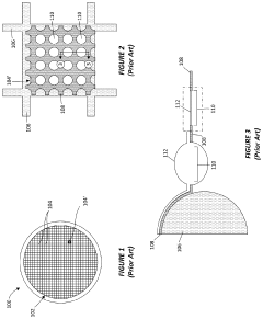
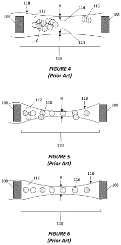
System and method for preparing cryo-em grids
PatentActiveUS20200303162A1
Innovation
- A cryogenically-cooled sample preparation apparatus with a movable and rotatable sample dispenser that automatically deposits and vitrifies liquid samples on a grid, eliminating the need for manual handling and blotting, and allowing for precise control of ice layer thickness through adjustable deposition rates and cryogen circulation.
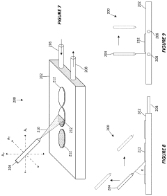
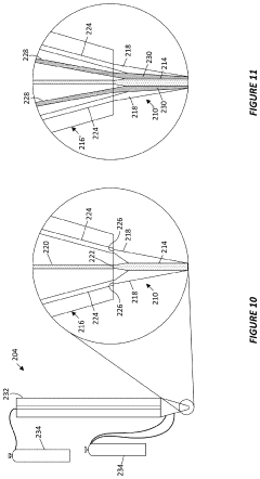
Cryo-em sample preparation method and apparatus
PatentWO2024189350A1
Innovation
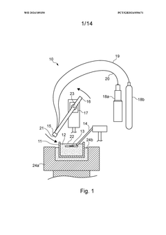
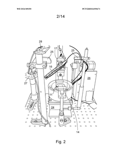
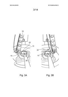
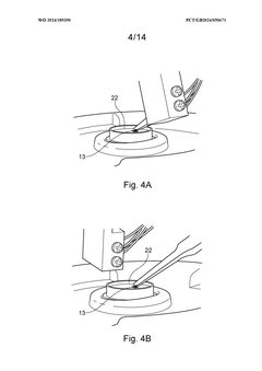
- A system that rapidly deposits a target analyte as a stream of sample droplets onto a cryogenically cooled substrate support, minimizing the time between deposition and vitrification to achieve submillisecond timescales, using a fluidic aerosol delivery system and cryogenic fluids to create a thin, uniform ice layer.
Environmental Impact and Sustainability Considerations
The environmental impact of cryo-electron microscopy (cryo-EM) for imaging liquid-containing nanomaterials extends beyond its scientific applications. The technique's sustainability profile must be evaluated comprehensively as its adoption increases across research and industrial sectors.
Energy consumption represents a significant environmental consideration in cryo-EM operations. The equipment requires substantial power for maintaining ultra-low temperatures (typically below -150°C) and operating high-voltage electron beams. A single cryo-EM facility can consume electricity equivalent to dozens of households, raising questions about carbon footprint in regions dependent on fossil fuel energy sources.
Liquid nitrogen and helium usage presents another environmental challenge. These cryogens are essential for sample vitrification and microscope operation, but their production involves energy-intensive air separation processes. Additionally, helium is a finite resource with limited global reserves, making its extensive use in imaging technologies potentially problematic from a resource conservation perspective.
Chemical waste management constitutes an important sustainability consideration. Sample preparation for liquid-containing nanomaterials often involves surfactants, heavy metals for contrast enhancement, and various organic solvents. Proper disposal protocols must be implemented to prevent environmental contamination, particularly as these materials may contain engineered nanoparticles with uncertain environmental fate and toxicity profiles.
Water usage in cryo-EM facilities, particularly for cooling systems, represents another environmental impact vector. Advanced facilities can consume thousands of gallons daily, necessitating water recycling systems to minimize consumption in water-stressed regions.
The lifecycle assessment of cryo-EM equipment reveals additional sustainability concerns. The manufacturing process involves rare earth elements and specialized materials with complex supply chains and extraction impacts. Furthermore, the limited lifespan of certain components (typically 5-10 years for major systems) creates electronic waste management challenges.
Encouragingly, technological innovations are addressing these environmental concerns. Energy-efficient microscope designs, improved thermal insulation, and automated imaging processes that optimize cryogen usage are reducing the environmental footprint. Additionally, shared facility models maximize equipment utilization rates, distributing the environmental impact across multiple research projects and reducing redundant infrastructure development.
Data Processing and Computational Analysis Advancements
The evolution of data processing and computational analysis in cryo-electron microscopy (cryo-EM) has been pivotal for advancing liquid-containing nanomaterial imaging. Traditional image processing algorithms struggled with the unique challenges posed by liquid samples, including lower signal-to-noise ratios and increased beam sensitivity. Recent advancements in machine learning and artificial intelligence have revolutionized how researchers extract meaningful structural information from these complex specimens.
Deep learning frameworks specifically designed for cryo-EM data have emerged as game-changers in the field. These algorithms can effectively distinguish between the signal from the nanomaterial structure and the noise from the surrounding liquid environment. Convolutional neural networks (CNNs) have demonstrated particular efficacy in automated particle picking and classification, reducing human bias and accelerating analysis workflows by orders of magnitude compared to manual methods.
Motion correction algorithms have seen significant refinement to address the unique challenges of liquid-phase samples. Beam-induced motion is more pronounced in liquid environments, requiring sophisticated computational approaches that can track and compensate for these movements at the sub-pixel level. Software packages like MotionCor2 and Relion have been adapted specifically for liquid-phase cryo-EM, incorporating models that account for the fluid dynamics within the sample.
3D reconstruction techniques have evolved to handle the heterogeneity inherent in liquid-containing nanomaterials. Maximum likelihood methods and Bayesian approaches now allow researchers to identify and classify multiple conformational states within a single dataset. This capability is crucial for understanding the dynamic behavior of nanomaterials in liquid environments, where structural flexibility often plays a key functional role.
Real-time processing capabilities represent another frontier in computational analysis for liquid-phase cryo-EM. On-the-fly data processing during image acquisition allows researchers to make informed decisions about imaging parameters and sample quality, maximizing the efficiency of microscope time and improving overall data quality. These systems leverage high-performance computing clusters and GPU acceleration to process the massive datasets generated during cryo-EM sessions.
Integration of molecular dynamics simulations with cryo-EM data has enhanced our understanding of nanomaterial-liquid interactions. By combining experimental imaging data with computational models, researchers can now visualize dynamic processes such as ligand binding, nanoparticle aggregation, and surface interactions with unprecedented detail. This hybrid approach bridges the gap between static structural information and dynamic functional insights.
Unlock deeper insights with Patsnap Eureka Quick Research — get a full tech report to explore trends and direct your research. Try now!
Generate Your Research Report Instantly with AI Agent
Supercharge your innovation with Patsnap Eureka AI Agent Platform!
