

Cryo-EM For Nanoparticle Surface Ligand Mapping
AUG 27, 20259 MIN READ
Generate Your Research Report Instantly with AI Agent
PatSnap Eureka helps you evaluate technical feasibility & market potential.
Cryo-EM Ligand Mapping Background and Objectives
Cryo-electron microscopy (Cryo-EM) has emerged as a revolutionary technique in structural biology, enabling researchers to visualize biological macromolecules at near-atomic resolution. In recent years, this powerful imaging method has been increasingly applied to the field of nanomaterials, particularly for mapping surface ligands on nanoparticles. The evolution of Cryo-EM technology has been marked by significant breakthroughs, from its initial development in the 1980s to the "resolution revolution" in the 2010s that earned Jacques Dubochet, Joachim Frank, and Richard Henderson the 2017 Nobel Prize in Chemistry.
Surface ligands play a crucial role in determining nanoparticle properties, including stability, solubility, biocompatibility, and functionality. Traditional characterization methods such as nuclear magnetic resonance (NMR), infrared spectroscopy, and mass spectrometry provide valuable information about ligand composition but often lack spatial resolution and cannot directly visualize ligand distribution on nanoparticle surfaces. This technological gap has hindered the rational design of nanoparticles for various applications in medicine, catalysis, and materials science.
The application of Cryo-EM to nanoparticle ligand mapping represents a convergence of structural biology techniques with nanomaterial characterization. This interdisciplinary approach has gained momentum as researchers recognize the potential to obtain unprecedented insights into ligand-nanoparticle interactions at the molecular level. The trend is moving toward higher resolution imaging, improved sample preparation techniques, and advanced computational methods for image processing and reconstruction.
The primary objective of Cryo-EM ligand mapping is to develop robust methodologies for visualizing and characterizing the spatial arrangement, conformation, and dynamics of surface ligands on nanoparticles with nanometer or sub-nanometer resolution. This includes optimizing sample preparation protocols to preserve the native state of ligands, enhancing contrast for visualization of organic molecules, and developing analytical frameworks for quantitative analysis of ligand distribution patterns.
Secondary objectives include correlating ligand architecture with nanoparticle properties and functions, enabling rational design of nanoparticles with tailored surface chemistry, and establishing standardized approaches for quality control in nanoparticle synthesis. The field also aims to integrate Cryo-EM data with complementary techniques such as molecular dynamics simulations and spectroscopic methods to create comprehensive models of nanoparticle surface chemistry.
Ultimately, the technological goal is to transition Cryo-EM from a specialized research tool to a routine analytical method for nanoparticle characterization in both academic and industrial settings, supporting innovation in nanomedicine, catalysis, energy storage, and advanced materials development.
Market Applications for Nanoparticle Surface Characterization
The market for nanoparticle surface characterization technologies is experiencing robust growth, driven by expanding applications across multiple industries. Pharmaceutical and biomedical sectors represent the largest market segment, where precise surface ligand mapping enables targeted drug delivery systems, improved biocompatibility, and enhanced therapeutic efficacy. The ability to visualize and analyze ligand distribution on nanoparticle surfaces using Cryo-EM provides critical insights for developing more effective nanomedicines and reducing side effects.
Materials science and advanced manufacturing constitute another significant market, where nanoparticle surface properties directly influence material performance. Industries developing catalysts, coatings, and composite materials benefit substantially from accurate surface characterization to optimize product functionality. The semiconductor industry also relies heavily on these technologies for quality control in nanoscale components and devices, where even minor surface irregularities can impact performance.
Environmental monitoring and remediation represent emerging application areas with substantial growth potential. Nanoparticles used in environmental sensing, water purification, and pollution control require precise surface engineering, making Cryo-EM-based characterization increasingly valuable. The food and agriculture sectors are also adopting these technologies for developing improved food packaging, agricultural inputs, and quality control measures.
The energy sector presents particularly promising opportunities, especially in battery technology and renewable energy applications. Surface ligand mapping helps optimize electrode materials, improve energy storage capacity, and enhance the efficiency of solar cells and fuel cells. As the global push for sustainable energy solutions intensifies, demand for advanced nanoparticle characterization is expected to grow significantly.
Consumer products incorporating engineered nanomaterials, from cosmetics to textiles, represent another expanding market segment. Manufacturers increasingly need sophisticated surface analysis to ensure product safety, efficacy, and regulatory compliance. The cosmetics industry, in particular, has seen growing demand for nanoparticle-based formulations with precisely engineered surface properties.
Academic and research institutions form a stable market base, consistently investing in advanced characterization technologies to support fundamental and applied research. Government laboratories focused on defense, security, and public health also represent significant customers for these technologies, particularly for applications in biosensing, threat detection, and protective materials.
Geographically, North America and Europe currently lead in market adoption, but the Asia-Pacific region is experiencing the fastest growth rate, driven by expanding research infrastructure and industrial development in China, Japan, South Korea, and India.
Current Limitations in Nanoparticle Ligand Visualization
Despite significant advancements in cryo-electron microscopy (cryo-EM) for nanoparticle characterization, several critical limitations persist in the visualization and mapping of surface ligands. The primary challenge stems from the inherent contrast limitations, as organic ligands typically consist of light elements (C, H, N, O) that scatter electrons weakly compared to the metallic or inorganic nanoparticle cores. This fundamental contrast disparity makes direct visualization of the ligand shell extremely difficult, particularly when attempting to resolve individual ligand molecules.
Resolution constraints further complicate ligand mapping efforts. While modern cryo-EM instruments can achieve sub-2Å resolution for protein structures, the dynamic and flexible nature of surface ligands often results in significant motion blur. This mobility reduces the effective resolution achievable for ligand visualization, frequently limiting detailed structural information about ligand conformation and organization on the nanoparticle surface.
Sample preparation introduces additional complications that impact visualization quality. The vitrification process central to cryo-EM can potentially alter the native state of ligand shells through ice formation or interaction with the air-water interface. Furthermore, beam damage from electron exposure progressively destroys organic ligands during imaging, creating a fundamental trade-off between obtaining sufficient signal and preserving the specimen's structural integrity.
Data processing challenges compound these physical limitations. Current computational approaches struggle to effectively distinguish between closely packed ligands or to accurately model their conformational heterogeneity. The lack of specialized algorithms for ligand-specific reconstruction represents a significant bottleneck in extracting meaningful information from cryo-EM datasets of functionalized nanoparticles.
Validation methods for ligand mapping results remain underdeveloped compared to those established for protein structures. The absence of standardized benchmarks and complementary techniques specifically designed to verify ligand arrangements creates uncertainty in data interpretation and limits confidence in structural models derived from cryo-EM observations.
Integration with other analytical techniques presents another limitation. While combining cryo-EM with spectroscopic methods (NMR, FTIR) or scattering techniques (SAXS) could potentially provide more comprehensive characterization, effective workflows for correlative analysis remain largely undeveloped, preventing researchers from fully leveraging complementary data sources to overcome the inherent limitations of any single technique.
Current Methodologies for Nanoparticle Surface Analysis
01 Cryo-EM techniques for surface ligand identification
Cryo-electron microscopy provides high-resolution imaging capabilities for identifying and mapping surface ligands on biological structures. These techniques allow researchers to visualize the binding sites and conformational changes of ligands on protein surfaces without the need for crystallization. Advanced image processing algorithms help enhance the contrast between ligands and their binding surfaces, enabling precise mapping of interaction interfaces at near-atomic resolution.- Cryo-EM techniques for surface ligand identification: Cryo-electron microscopy provides high-resolution imaging capabilities for identifying and mapping surface ligands on biological structures. These techniques allow researchers to visualize the binding sites and interactions between ligands and their target molecules at near-atomic resolution. By flash-freezing samples in their native state, cryo-EM preserves the structural integrity of ligand-receptor complexes, enabling accurate mapping of surface ligands without the need for crystallization.
- Integration of computational methods with cryo-EM for ligand mapping: Advanced computational algorithms and software tools are integrated with cryo-EM data to enhance surface ligand mapping. These computational methods include image processing, 3D reconstruction, molecular dynamics simulations, and machine learning approaches that help interpret cryo-EM density maps and identify ligand binding sites. The combination of experimental cryo-EM data with computational analysis improves the accuracy and efficiency of surface ligand identification and characterization.
- Cryo-EM for drug discovery and development: Cryo-EM surface ligand mapping plays a crucial role in drug discovery and development by providing detailed structural information about drug-target interactions. This approach enables researchers to visualize how potential therapeutic compounds bind to their targets, facilitating structure-based drug design and optimization. The high-resolution structural data obtained through cryo-EM helps in understanding the mechanism of action of drugs and can lead to the development of more effective and selective therapeutic agents.
- Sample preparation techniques for cryo-EM ligand mapping: Specialized sample preparation methods are essential for successful cryo-EM surface ligand mapping. These techniques include grid preparation, vitrification processes, and specific protocols for preserving ligand-receptor complexes in their native state. Innovations in sample preparation, such as the use of graphene supports, specialized detergents, and optimized buffer conditions, have significantly improved the resolution and quality of cryo-EM data for ligand mapping applications.
- Cryo-EM for characterizing nanoparticle-ligand interactions: Cryo-EM is increasingly used to characterize the surface properties and ligand distribution on nanoparticles used in various applications, including drug delivery systems and diagnostic agents. This technique allows for direct visualization of ligand density, orientation, and conformation on nanoparticle surfaces, providing critical information for optimizing nanoparticle design and functionality. The ability to map ligands on nanoparticle surfaces helps in understanding their biological interactions and improving their targeting efficiency.
02 Integration of computational methods with Cryo-EM for ligand mapping
Computational approaches combined with Cryo-EM data enhance the accuracy of surface ligand mapping. Machine learning algorithms and molecular dynamics simulations help interpret electron density maps to identify ligand binding sites. These computational methods can predict ligand-receptor interactions, optimize molecular docking, and validate structural models derived from Cryo-EM data, providing comprehensive insights into the spatial arrangement of ligands on molecular surfaces.Expand Specific Solutions03 Sample preparation innovations for Cryo-EM ligand mapping
Novel sample preparation methods improve the visualization of surface ligands in Cryo-EM. Techniques such as vitrification optimization, grid functionalization, and contrast enhancement agents help preserve the native state of ligand-receptor complexes. These innovations reduce sample damage during preparation, enhance signal-to-noise ratio, and maintain the structural integrity of delicate surface ligands, resulting in more accurate mapping of ligand positions on molecular surfaces.Expand Specific Solutions04 Cryo-EM for mapping antibody and therapeutic ligand interactions
Cryo-EM enables detailed mapping of therapeutic antibodies and drug candidates binding to target surfaces. This application is particularly valuable for epitope mapping, characterizing antibody-antigen interfaces, and optimizing drug design. The technique allows visualization of conformational epitopes that are difficult to identify using other methods, providing crucial information for vaccine development and therapeutic antibody engineering by revealing the precise binding mechanisms of ligands to their targets.Expand Specific Solutions05 Time-resolved Cryo-EM for dynamic ligand mapping
Time-resolved Cryo-EM techniques capture the dynamic nature of ligand-receptor interactions on molecular surfaces. By freezing samples at different time points after ligand introduction, researchers can observe binding kinetics, conformational changes, and transient interactions. This approach provides insights into the temporal aspects of ligand recognition and binding, revealing intermediate states and structural rearrangements that occur during the binding process, which is crucial for understanding the functional mechanisms of molecular interactions.Expand Specific Solutions
Leading Research Groups and Commercial Entities in Cryo-EM
Cryo-EM for nanoparticle surface ligand mapping is currently in a growth phase, with the market expanding as this technique addresses critical challenges in nanomaterial characterization. The global market is estimated to reach several hundred million dollars by 2025, driven by pharmaceutical and materials science applications. Technologically, the field is advancing rapidly but remains specialized, with key players demonstrating varying levels of expertise. Leading research institutions like The Rockefeller University, Max Planck Society, and Tsinghua University are pioneering fundamental research, while commercial entities such as FEI Co. (microscopy equipment), Quantifoil Micro Tools (specialized grids), and DENSsolutions (in-situ holders) are developing enabling technologies. Emerging companies like Ion Dx and MiTeGen are introducing innovative sample preparation solutions, indicating a maturing ecosystem with opportunities for specialized applications.
FEI Co.
Technical Solution: FEI Co. (now part of Thermo Fisher Scientific) has developed advanced cryo-EM technology specifically optimized for nanoparticle surface ligand mapping. Their Titan Krios platform incorporates direct electron detectors and phase plates that enable visualization of surface ligands with near-atomic resolution. The company's proprietary image processing algorithms enhance contrast between ligands and nanoparticle cores, allowing researchers to distinguish between different ligand types and their spatial distribution[1]. FEI's technology includes specialized sample preparation techniques that preserve the native state of ligand shells during vitrification, minimizing artifacts that could distort ligand arrangement. Their integrated workflow combines hardware innovations with software solutions that perform 3D reconstruction of ligand shells, providing quantitative data on ligand density, orientation, and conformation around nanoparticles[3]. Recent advancements include energy-filtered imaging capabilities that improve signal-to-noise ratios specifically for organic ligands with low atomic numbers.
Strengths: Industry-leading resolution capabilities allowing visualization of even small organic ligands; comprehensive integrated workflow from sample preparation to 3D reconstruction; extensive global support network. Weaknesses: High equipment costs limiting accessibility; requires significant expertise for optimal operation; sample preparation remains challenging for certain nanoparticle-ligand combinations.
President & Fellows of Harvard College
Technical Solution: Harvard University researchers have developed a sophisticated cryo-EM approach for nanoparticle surface ligand mapping that combines advanced imaging techniques with computational modeling. Their methodology employs specialized vitrification protocols that minimize ice crystal formation and preserve the native conformation of surface ligands[2]. Harvard's approach incorporates energy-filtered transmission electron microscopy (EFTEM) to enhance contrast between organic ligands and inorganic nanoparticle cores, allowing for improved visualization of ligand shells. The technology utilizes custom image processing algorithms that can identify and track individual ligand molecules on nanoparticle surfaces, enabling quantitative analysis of ligand density, distribution patterns, and conformational states[4]. Harvard researchers have pioneered the integration of molecular dynamics simulations with experimental cryo-EM data, creating hybrid models that provide insights into ligand-ligand interactions and ligand-solvent dynamics. Their platform also includes correlative techniques that combine cryo-EM with spectroscopic methods such as EELS (Electron Energy Loss Spectroscopy) to provide chemical fingerprinting of different ligand types on the same nanoparticle[8].
Strengths: Cutting-edge integration of experimental and computational approaches; ability to provide dynamic information about ligand behavior; high-resolution capabilities for complex nanoparticle systems. Weaknesses: Highly specialized expertise required; techniques not yet widely accessible outside research environment; computational modeling aspects require validation through multiple experimental approaches.
Key Technical Advances in Cryo-EM Ligand Resolution
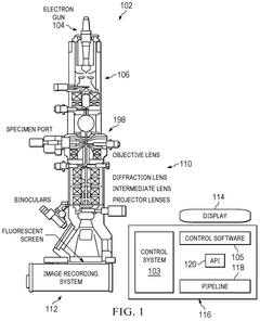
Automating cryo-electron microscopy data collection
PatentWO2024229329A1
Innovation
- A software pipeline utilizing machine learning models for automated navigation of cryo-EM grids, including square and hole localization, scoring, and on-the-fly learning, to determine high-quality targeting locations without human input, using pretrained models and active learning techniques like Gaussian Process regression.

Computational Methods for Ligand Density Reconstruction
Computational methods for ligand density reconstruction represent a critical component in the analysis of cryo-electron microscopy (cryo-EM) data for nanoparticle surface ligand mapping. These methods bridge the gap between raw experimental data and meaningful structural insights about ligand organization on nanoparticle surfaces.
The reconstruction process typically begins with image processing algorithms that enhance signal-to-noise ratio in cryo-EM micrographs. Techniques such as contrast transfer function (CTF) correction, motion correction, and particle picking are essential preprocessing steps before ligand density can be accurately reconstructed. Advanced denoising algorithms, including wavelet-based methods and deep learning approaches, have shown particular promise in extracting meaningful signal from the inherently noisy cryo-EM data.
Following preprocessing, 3D reconstruction algorithms transform 2D projections into volumetric representations. For ligand mapping specifically, subtraction methods have proven valuable, where the known core nanoparticle density is computationally removed to isolate the ligand shell contribution. This approach enhances the visibility of the often lower-density ligand structures surrounding the high-density nanoparticle core.
Machine learning techniques have revolutionized ligand density reconstruction in recent years. Convolutional neural networks (CNNs) trained on simulated cryo-EM data can identify subtle patterns in experimental images that traditional algorithms might miss. Generative adversarial networks (GANs) have demonstrated capability in predicting missing information in sparse datasets, potentially addressing the limited-angle problem inherent in many cryo-EM experiments.
Molecular dynamics (MD) simulations serve as complementary computational tools, providing physically realistic constraints for ligand conformations. By integrating MD simulations with experimental cryo-EM densities, researchers can generate hybrid models that satisfy both experimental data and theoretical energy minimization principles. This integration often employs Bayesian statistical frameworks to weight the contributions of experimental and theoretical inputs appropriately.
Recent advances include the development of specialized software packages that incorporate these computational methods into user-friendly workflows. Programs such as RELION, cryoSPARC, and EMAN2 have implemented modules specifically designed for heterogeneous reconstruction problems like ligand mapping. These tools increasingly incorporate GPU acceleration to handle the computational demands of processing large cryo-EM datasets.
The field continues to evolve toward higher resolution reconstructions and improved discrimination between different ligand types on the same nanoparticle surface. Emerging computational approaches focus on identifying dynamic behaviors of ligands rather than static structures, providing insights into the functional properties of nanoparticle-ligand systems.
Sample Preparation Challenges and Innovations
Sample preparation represents one of the most critical and challenging aspects of cryo-electron microscopy (cryo-EM) for nanoparticle surface ligand mapping. The quality of structural data obtained is directly dependent on the preparation techniques employed, with several persistent challenges requiring innovative solutions.
The primary challenge in sample preparation stems from the delicate nature of the ligand-nanoparticle interface. Surface ligands are often dynamic, flexible structures that can be easily disrupted during conventional preparation procedures. The vitrification process, while essential for preserving native structures, can introduce artifacts if not optimized specifically for ligand-coated nanoparticles. Parameters such as blotting force, humidity, and temperature must be precisely controlled to maintain ligand integrity.
Contrast generation presents another significant hurdle. Many organic ligands exhibit poor electron contrast against the vitreous ice background, making them difficult to visualize. This challenge is particularly pronounced for small ligands or those with low electron density. Researchers have addressed this through selective staining approaches and the development of high-contrast ligand analogs that maintain similar binding properties while offering improved visibility.
Heterogeneity in ligand distribution across nanoparticle surfaces complicates comprehensive mapping efforts. Traditional preparation methods may inadvertently select for certain particle orientations at the air-water interface, resulting in incomplete structural information. Recent innovations include the use of graphene oxide or functionalized carbon films as support substrates to minimize preferential orientation effects.
Cryo-EM grid preparation has seen significant advancements specifically tailored for nanoparticle systems. The introduction of self-wicking grids has reduced blotting-induced artifacts, while specialized plasma cleaning protocols help optimize surface hydrophilicity for more uniform sample distribution. Microfluidic approaches for sample deposition have emerged as promising alternatives to traditional blotting methods, offering more gentle handling of delicate ligand structures.
The development of correlative techniques has further enhanced sample preparation capabilities. By combining cryo-EM with complementary methods such as super-resolution fluorescence microscopy, researchers can now identify regions of interest before detailed structural analysis, improving efficiency and targeting specific ligand arrangements on nanoparticle surfaces.
Cryogenic focused ion beam (cryo-FIB) milling has revolutionized sample preparation for complex nanoparticle assemblies, allowing precise thinning of vitrified samples to ideal thicknesses for transmission electron microscopy while preserving the native state of surface ligands. This approach has proven particularly valuable for studying nanoparticle interactions in more complex environments that better mimic physiological conditions.
Unlock deeper insights with PatSnap Eureka Quick Research — get a full tech report to explore trends and direct your research. Try now!
Generate Your Research Report Instantly with AI Agent
Supercharge your innovation with PatSnap Eureka AI Agent Platform!