Two-dimensional medical image tissue and organ pseudo color display method and device

A technology of tissues and organs and medical images, which is applied in the field of medical imaging, can solve the problems that multiple tissues and organs cannot be displayed in false colors, false color images overlap each other, and the display is not clear, etc., to achieve simple display effects, avoid mutual overlap, and avoid color mixing Effect

Active Publication Date: 2016-11-23
QINGDAO HISENSE MEDICAL EQUIP
View PDF5 Cites 3 Cited by
  • Summary
  • Abstract
  • Description
  • Claims
  • Application Information

AI Technical Summary

Problems solved by technology

[0004] The existing technology can only perform pseudo-color display on a single organ on a two-dimensional medical image, but cannot perform pseudo-color display on multiple tissues and organs at the same time, and the flexibility is low
Moreover, since two-dimensional medical images may include multiple organs, and each organ may include multiple tissues, such as blood vessels, tumors, etc., if multiple tissues and organs are displayed in pseudo-color according to the above-mentioned single-organ pseudo-color display method , there may be overlapping of false color images between organs and organs, between organs and tissues, or between tissues, resulting in unclear display

Method used


Image

  • Two-dimensional medical image tissue and organ pseudo color display method and device
  • Two-dimensional medical image tissue and organ pseudo color display method and device
  • Two-dimensional medical image tissue and organ pseudo color display method and device

Examples


Embodiment Construction

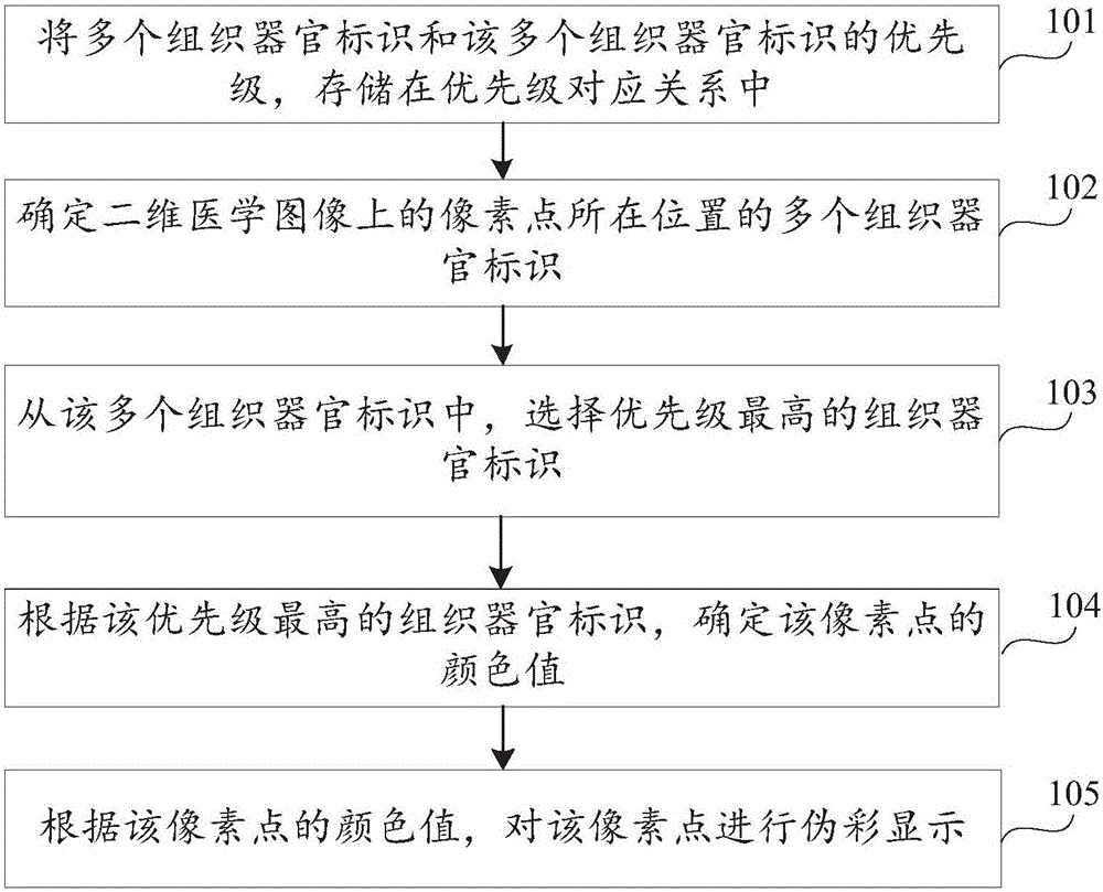
[0056] In order to make the object, technical solution and advantages of the present invention clearer, the implementation manner of the present invention will be further described in detail below in conjunction with the accompanying drawings.

[0057] Before explaining and describing the embodiments of the present invention in detail, the application scenarios of the embodiments of the present invention are firstly introduced. The method provided by the embodiment of the present invention is applied to a terminal, and the terminal can be a computer, a smart phone, a tablet computer, a notebook computer, an ultra-mobile personal computer (English: Ultra-mobile Personal Computer, UMPC for short), a netbook, a personal digital assistant (English: Personal Digital Assistant, PDA for short), etc., may also be a two-dimensional medical image acquisition device such as a CT machine or a nuclear magnetic resonance apparatus, which is not limited in the embodiment of the present invent...


PUM


Abstract

The invention discloses a two-dimensional medical image tissue and organ pseudo color display method, and belongs to the field of medical images. The method includes the steps that multiple tissue and organ identifications of the position, where a pixel point is located, on a two-dimensional medical image are determined, and each of the tissue and organ identifications comprises a tissue identification body and an organ identification body; the tissue and organ identification with the highest priority is selected from the tissue and organ identifications; according to the tissue and organ identification with the highest priority, the color value of the pixel point is determined; the pixel point is displayed in a pseudo color mode according to the color value of the pixel point. The pixel point with the position, corresponding to the tissue and organ identifications, on the two-dimensional medical image is displayed in the pseudo color mode according to the color value indicated by the tissue and organ identification having the highest priority and located at the position where the pixel point is located, color mixture of multiple color values in the pixel point is avoided, pseudo color image overlap among the multiple organs or tissue is avoided, the display effect is concise and clear, and flexibility is high.

Description

technical field [0001] Embodiments of the present invention relate to the field of medical imaging, and in particular to a method and device for displaying tissues and organs in pseudo-color in two-dimensional medical images. Background technique [0002] In recent years, with the development of medical imaging technology, two-dimensional medical images obtained by medical imaging technologies such as CT (Computed Tomography, computerized tomography) and MRI (Magnetic Resonance Imaging, magnetic resonance imaging) have been more and more clinically applied. more extensive. Among them, the structural information of multiple tissues and organs can be intuitively displayed on the two-dimensional medical image, which can assist doctors in observation and clinical diagnosis, and each of the multiple tissues and organs can be a tissue or an organ, etc., but due to The current two-dimensional medical images are usually displayed in gray scale, and the display of various tissues an...

Claims


Application Information

Patent Timeline
Patent Type & Authority Applications(China)
IPC IPC(8): G06F19/00G06T7/00
CPCG06F19/321G06T2207/10081G06T2207/30004
Inventor 田广野陈哲吴海燕
Owner QINGDAO HISENSE MEDICAL EQUIP
Why Eureka
Try Eureka