Visual 3D endoscope capable of guiding tracheal intubation

A technology of endotracheal intubation and endoscopy, which is applied in the field of medical equipment, can solve the problems of large physical injuries, many limiting factors, inability to intubate in patients with difficult airways and emergency airways, and achieve easy operation and clear vision , the effect of solving difficult airway and emergency airway

Pending Publication Date: 2017-12-26
高长胜
View PDF5 Cites 8 Cited by
  • Summary
  • Abstract
  • Description
  • Claims
  • Application Information

AI Technical Summary

Problems solved by technology

[0006] To sum up, the problems existing in the prior art are: laryngoscopes, video laryngoscopes, light sticks, fiberoptic bronchoscopes, visual tube cores, and high-frequency ventilation on the market have many limiting factors and are subject to many external interferences. Excessive physical impairment precludes endotracheal tube intubation in patients with difficult and emergency airways

Method used


Image

  • Visual 3D endoscope capable of guiding tracheal intubation
  • Visual 3D endoscope capable of guiding tracheal intubation

Examples


Embodiment Construction

[0020] In order to further understand the content, features and effects of the present invention, the following examples are given, and detailed descriptions are given below with reference to the accompanying drawings.

[0021] The structure of the present invention will be described in detail below in conjunction with the accompanying drawings.

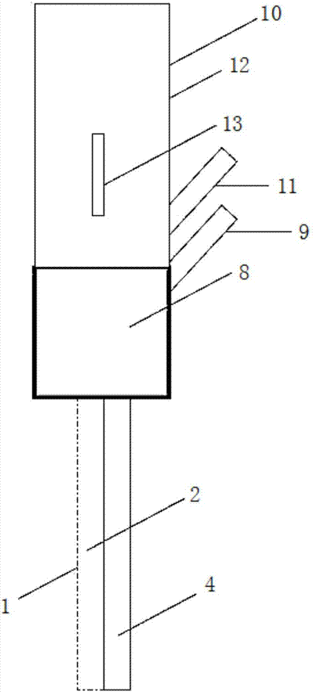
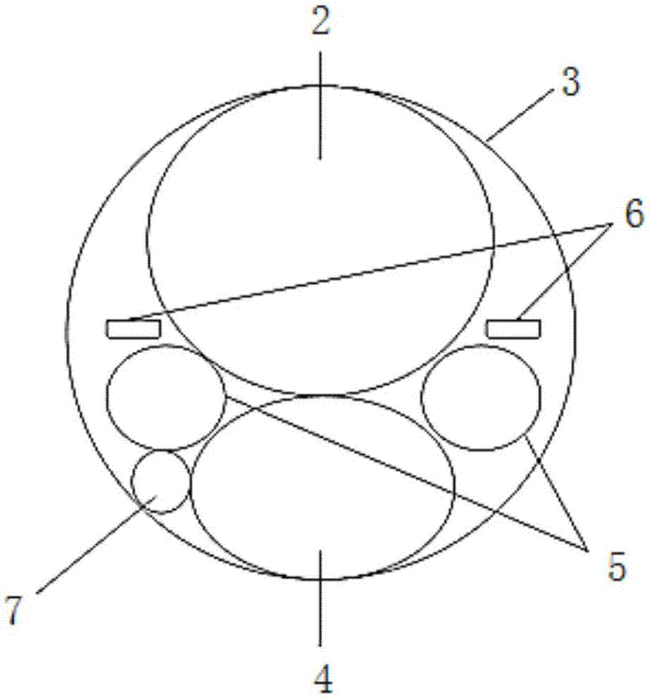
[0022] The visible 3D endoscope that can guide tracheal intubation is provided with a mirror body 1, and there is an exchange stylet guide groove 2 inside the back side of the scope body 1, and the exchange stylet guide groove 2 is in phase with the base 3 of the 3D endoscope. Connection; the ventral side of the mirror body 1 is provided with a punching hole 4, and the punching hole 4 is distributed on the opposite side of the guide groove 2 of the exchange die;

[0023] Two miniature cameras 5, two light sources 6 and a miniature microphone 7 are inlaid on the front end surface of the mirror body 1. On the side, two light sources 6...


PUM


Abstract

The invention belongs to the technical field of medical equipment, and discloses a visual 3D endoscope capable of guiding tracheal intubation. The visual 3D endoscope capable of guiding the tracheal intubation is provided with an endoscope body, wherein an exchange tube core guide groove is formed in the back side of the endoscope body, and the exchange tube core guide groove is connected with a base of the 3D endoscope; a sucking and flushing hole is formed in the ventral side of the endoscope body, and the sucking and flushing hole is distributed at the opposite side of the exchange tube core guide groove; two miniature cameras, two light sources and a micro microphone are inlaid on the front end surface of the endoscope body, the two miniature cameras are symmetrically arranged on two sides of the joint of the sucking and flushing hole and the exchange tube core guide groove, the two light sources are symmetrically distributed at the upper parts of the miniature cameras and the lower part of the exchange tube core guide groove, and the miniature microphone is arranged at the joint of the miniature camera on the left side and the sucking and flushing hole. The visual 3D endoscope capable of guiding the tracheal intubation integrates the current mainstream tracheal intubation devices and ideas including the laryngoscope, the visible laryngoscope, the light wand, the bronchofiberscope, the visual tube core, high-frequency ventilation and the like.

Description

technical field [0001] The invention belongs to the technical field of medical equipment, in particular to a visual 3D endoscope that can guide a tracheal intubation. Background technique [0002] The intubation fiberoptic bronchoscope and visual tube endoscope currently on the market have a small diameter because they must be placed inside the endotracheal tube, and cannot be equipped with more equipment to complete more functions. The field of view is narrow and the operation is cumbersome. One-time success rate, long time, fragile equipment, and easy to be disturbed by the suffix tongue base, oral secretions, vomit, etc. during the operation. [0003] Laryngoscopes, video laryngoscopes, etc. enter the oral cavity with a large volume, and the mouth opening is too small to be used, which is easy to damage the teeth; stimulate the epiglottis valle or the dorsal side of the epiglottis, cause greater side damage during operation, and the patient’s stress response is stronger; ...

Claims


Application Information

Patent Timeline
Patent Type & Authority Applications(China)
IPC IPC(8): A61B1/267A61B1/04A61B1/06A61M16/04
CPCA61M16/04A61B1/00193A61B1/04A61B1/06A61B1/267
Inventor 高长胜黄符香
Owner 高长胜
Why Eureka
Try Eureka