Method, computer and imaging apparatus for evaluating medical image data of an examination subject

a technology of medical image data and computer, applied in the field of method for evaluating medical image data of an examination subject, can solve problems such as inaccessibility of data, and achieve the effect of increasing the sensitivity of an evaluation and increasing the sensitivity

Active Publication Date: 2017-05-11
SIEMENS HEALTHCARE GMBH
View PDF6 Cites 3 Cited by
  • Summary
  • Abstract
  • Description
  • Claims
  • Application Information

AI Technical Summary

Benefits of technology

[0017]The inventive approach is accordingly based on the combined use of medical imaging and diagnostic methods, for example molecular diagnostics, which serve to acquire the clinical marker of the examination subject. With the inventive approach it is possible to determine normal value ranges for the physiological parameter that are individually matched to the examination subject. Thus, it is possible to increase the sensitivity of an evaluation of the medical image data in relation to the value of the physiological parameter determined on the basis of the medical image data. The diagnostic value of the measured value of the physiological parameter can also be increased in this way.
[0018]In an embodiment, genetic data of the examination subject are acquired as the clinical marker. The genetic data can be acquired by a molecular diagnostic method known to those skilled in the art. Genetic data relates to the hereditary characteristics specific to the examination subject, for example a part of a DNA sequence of the examination subject or a part of the genetic fingerprint of the examination subject. The genetic data of the examination subject that are acquired have an influence on the physiological parameter that is quantified on the basis of the medical image data. The genetic data actually acquired from the examination subject can therefore depend on the clinical problem. The status of the examination subject that is characterized by the genetic data can then be the presence of a gene variant in the examination subject or a change in a DNA section in a region between genes (intergenic variant). The status of the examination subject identified in this way on the basis of the genetic data can then be used to establish the normal value range for the physiological parameter that is matched to the status of the examination subject. This approach is based on the consideration that the genetic data of the examination subject can have an effect on a value range in which the values of the physiological parameter can be regarded as physiological and innocuous. In the case of an examination subject in which certain genetic data, for example a gene variant, is present, a measured value of the physiological parameter can point to the presence of a pathology, even though said measured value would be regarded as innocuous in relation to the standard normal value range for a larger patient population. Taking the genetic data into consideration in the adjustment of the normal value range can therefore lead to an increase in sensitivity in the assessment of the medical image data.
[0019]In another embodiment, genetic data are acquired that have an effect on a characteristic of an organ structure of the examination subject, wherein the physiological parameter reflects the characteristic of the organ structure. In this case the organ structure can be an organ of the examination subject, a delimited part of the examination subject, a group of organs of the examination subject, etc. The characteristic can in this case be a morphological or functional characteristic of the organ structure. The genetic data of the examination subject in this case has a physiological influence on the characteristic of the organ structure. The physiological parameter that reflects the characteristic of the organ structure must in this case be quantifiable on the basis of the medical image data.

Problems solved by technology

However, the presence of the reference image data is necessary for this approach, and such data are not available in most cases.

Method used


Image

  • Method, computer and imaging apparatus for evaluating medical image data of an examination subject
  • Method, computer and imaging apparatus for evaluating medical image data of an examination subject
  • Method, computer and imaging apparatus for evaluating medical image data of an examination subject

Examples


first embodiment

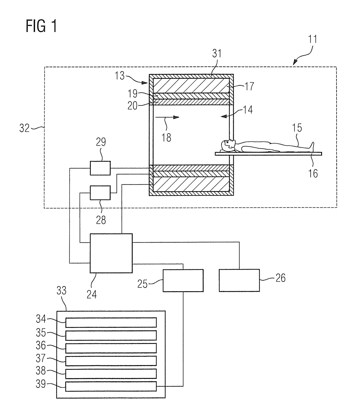
[0047]FIG. 2 shows a flowchart of the inventive method for evaluating medical image data.

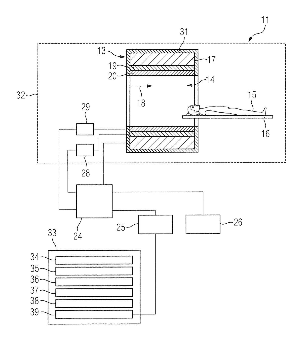
[0048]In a first method step 40, a clinical marker of the examination subject is acquired by means of the first acquisition processor 34, wherein the clinical marker characterizes a status of the examination subject in relation to a physiological parameter.

[0049]In a further method step 41, a normal value range for the physiological parameter that is matched to the status of the examination subject is ascertained as a function of the clinical marker by the ascertainment processor 35.

[0050]In a further method step 42, medical image data of the examination subject is acquired by the second acquisition processor 36.

[0051]In a further method step 43, a value of the physiological parameter of the examination subject is determined by means of the determination processor 37 using the medical image data.

[0052]In a further method step 44, the value of the physiological parameter is compared with the norm...

second embodiment

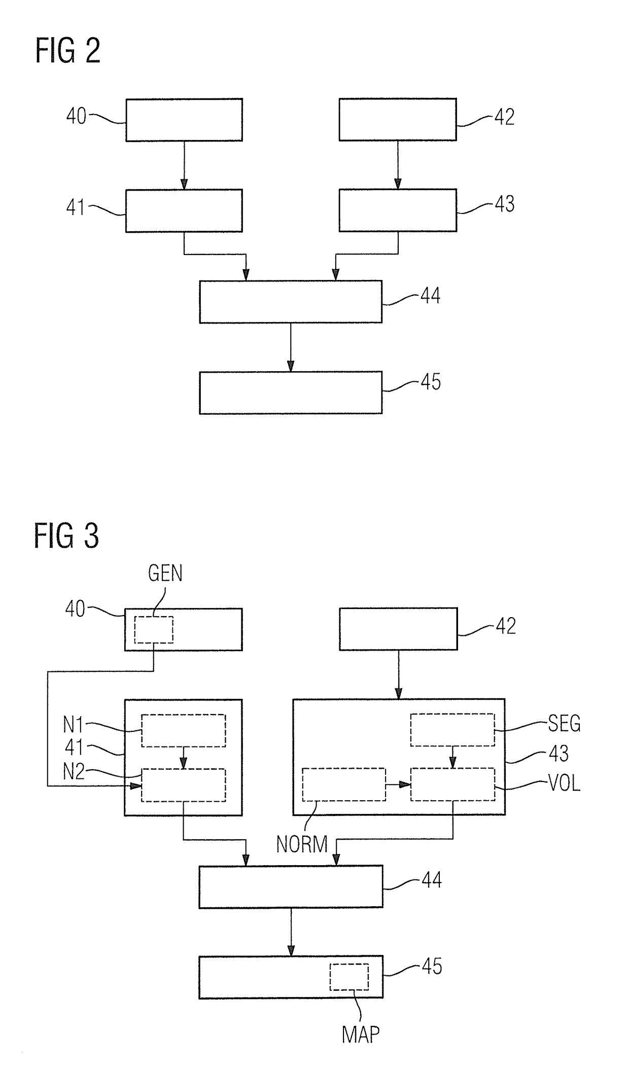
[0054]FIG. 3 shows a flowchart of the inventive method for evaluating medical image data.

[0055]The following description limits itself essentially to the differences compared to the exemplary embodiment in FIG. 2, with reference being made to the description of the exemplary embodiment in FIG. 2 in respect of method steps that remain the same. Method steps that remain substantially the same are labeled consistently with the same reference numerals.

[0056]The embodiment of the inventive method shown in FIG. 3 essentially includes the method steps 40, 41, 42, 43, 44, 45 of the first embodiment of the inventive method according to FIG. 2. The embodiment of the inventive method shown in FIG. 3 has additional method steps and substeps. An alternative method execution sequence to FIG. 3 that includes only some of the additional method steps and / or substeps shown in FIG. 3 is also conceivable. An alternative method execution sequence to FIG. 3 can of course also include additional method st...


PUM


Abstract

In a method and computer for evaluating medical image data of an examination subject, a clinical marker of the examination subject is acquired that characterizes a status of the examination subject in relation to a physiological parameter, and a normal value range for the physiological parameter is ascertained that is matched to the status of the examination subject as a function of the clinical marker. Medical image data of the examination subject are acquired, and a value of the physiological parameter of the examination subject is determined using the medical image data. This value is compared with the normal value range matched to the status of the examination subject, and the result of the comparison is provided as an output.

Description

BACKGROUND OF THE INVENTION[0001]Field of the Invention[0002]The invention concerns a method for evaluating medical image data of an examination subject, as well as an evaluation computer, a medical imaging apparatus and a non-transitory data storage medium for implementing such a method.[0003]Description of the Prior Art[0004]Typically, medical image data are recorded by medical imaging systems and can represent anatomical structures and / or functional processes of the body of an examination subject. An approach for evaluating the medical image data is known in which the medical image data acquired from the examination subject are compared with reference image data of the same examination subject in a healthy state, i.e. in particular in a state prior to the onset of disease symptoms. However, the presence of the reference image data is necessary for this approach, and such data are not available in most cases.[0005]A further possibility for evaluating the medical image data is know...

Claims


Application Information

Patent Timeline
Patent Type & Authority Applications(United States)
IPC IPC(8): G06K9/62G06T7/00A61B6/03A61B8/00A61B5/00A61B5/055
CPCG06K9/6202A61B5/72A61B5/055A61B6/037A61B6/032G06T2207/30016A61B5/0042A61B5/4088G06T7/0079G06T2207/30204A61B8/00A61B6/5217A61B8/5223G06T7/0012G06V10/751
Inventor FEIWEIER, THORSTENWELZEL, OLIVER PATRICK
Owner SIEMENS HEALTHCARE GMBH
Why Eureka
Try Eureka