Slit and slot scan, SAR, and compton devices and systems for radiation imaging

a technology of radiation imaging and compton devices, which is applied in the direction of instruments, radiation measurement, measurement devices, etc., can solve the problems of adding a degree of complexity to the assembling and maintenance of the detector system, multiple detector modules will have to be aligned with the x-ray beam, and the installation of a new detector array, so as to promote additional scatter reduction, reduce production and maintenance costs, and cost-effective

Inactive Publication Date: 2011-09-13
MINNESOTA IMAGING & ENG
View PDF16 Cites 36 Cited by
  • Summary
  • Abstract
  • Description
  • Claims
  • Application Information

AI Technical Summary

Benefits of technology

[0008]The invention utilizes the recent improvements in scintillators and photodetectors developed for nuclear medicine, high energy physics, etc. to develop cost-effective slit and slot scan detector systems for radiology with imaging properties comparable or superior to existing slit or slot scan detector systems. Energy integration, photon counting, and limited energy resolution detection modes can now be implemented whereas the current scintillator-based slit and slot scan systems implement energy integration only. In addition, changes in detector scan geometry can be employed to reduce production and maintenance costs while promoting additional scatter reduction. Although applications discussed herein are primarily directed at x-ray photon detection (which includes gamma ray photons for the purposes of this disclosure), in principle the invention can also be used to detect charged particles (alphas, betas, protons, etc.) and neutrons (as well as other neutral particles).
[0011]Slit and slot scan designs typically employ one or more slits or slots. The slit or slot length is significantly longer than the slit or slot width. The substantial length dimension often requires forming long arrays of scintillator materials (continuous or from discrete elements) that must be coupled to long photodetector arrays. Long photodetector arrays can consist of a single photodetector module but are often made by butting smaller photodetector array modules together. This adds a degree of complexity in assembling a detector system as well as in maintenance (the problem of repairing or replacing one or more dysfunctional readout elements. A similar problem exists when assembling a long photoemissive-based detector array. A single defect could require installing a new detector array and returning the old detector array to the factory. Typically, as the length dimension of the slit or slot increases (the area of the open aperture increases) the relative level of detected x-ray scatter increases. If two or more slits or slots are employed in a scanner they are offset so that they cover only part of the image during the scanning motion (avoiding scanning the same tissue volume multiple times) and reduce the overall scan time proportional to the number of slits or slots. While single- and multiple-slit or slot scanning geometries can be used with the detectors described in this invention, additional flexibility is introduced by dividing a slit or slot along the length dimension into sub-slits or sub-slots. The detector would be divided along the length dimension into detector modules to match the lengths of the sub-slits or sub-slots. A simple implementation of a scanning geometry is to have every other sub-slit or sub-slot and its corresponding detector module offset (staggered) from the original continuous slit or slot. This design would help reduce the relative level of detected x-ray scatter at each detector module. A large continuous detector can now be approximated by a set of detector modules that would be much less expensive to construction and repair (or replace). The design of focused detectors is simplified since individual modules can be optimized to compensate for local beam divergence. If the modules are sufficiently small, varying the tilt of modules according to their distance form the center of the beam is another method of compensating for beam divergence. A drawback to the offset detector geometry is that the multiple detector modules will have to be aligned with the x-ray beams. Depending on the application and the offset between sub-slits or sub-slots and their detector modules, the x-ray beams corresponding to certain detector modules may have to be blocked prior to and after exceeding, respectively, the desired scan area in order to avoid unnecessary dose to areas that need not be imaged.

Problems solved by technology

This adds a degree of complexity in assembling a detector system as well as in maintenance (the problem of repairing or replacing one or more dysfunctional readout elements.
A similar problem exists when assembling a long photoemissive-based detector array.
A single defect could require installing a new detector array and returning the old detector array to the factory.
A drawback to the offset detector geometry is that the multiple detector modules will have to be aligned with the x-ray beams.

Method used


Image

  • Slit and slot scan, SAR, and compton devices and systems for radiation imaging
  • Slit and slot scan, SAR, and compton devices and systems for radiation imaging
  • Slit and slot scan, SAR, and compton devices and systems for radiation imaging

Examples


Embodiment Construction

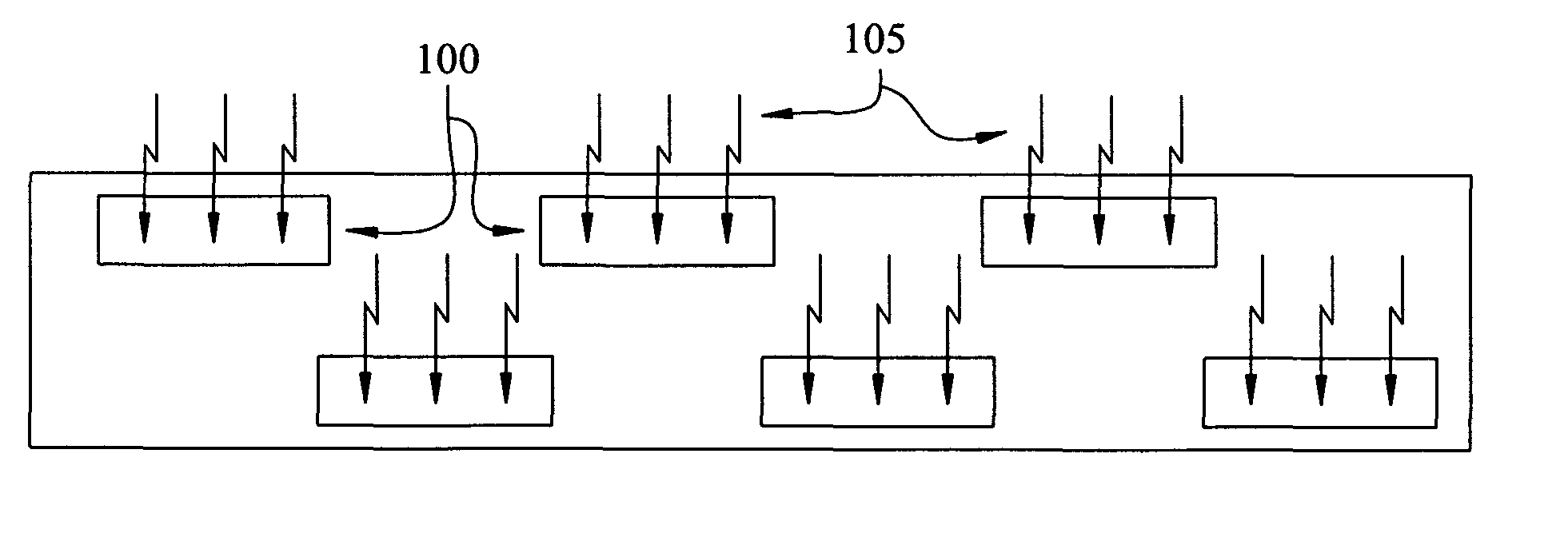
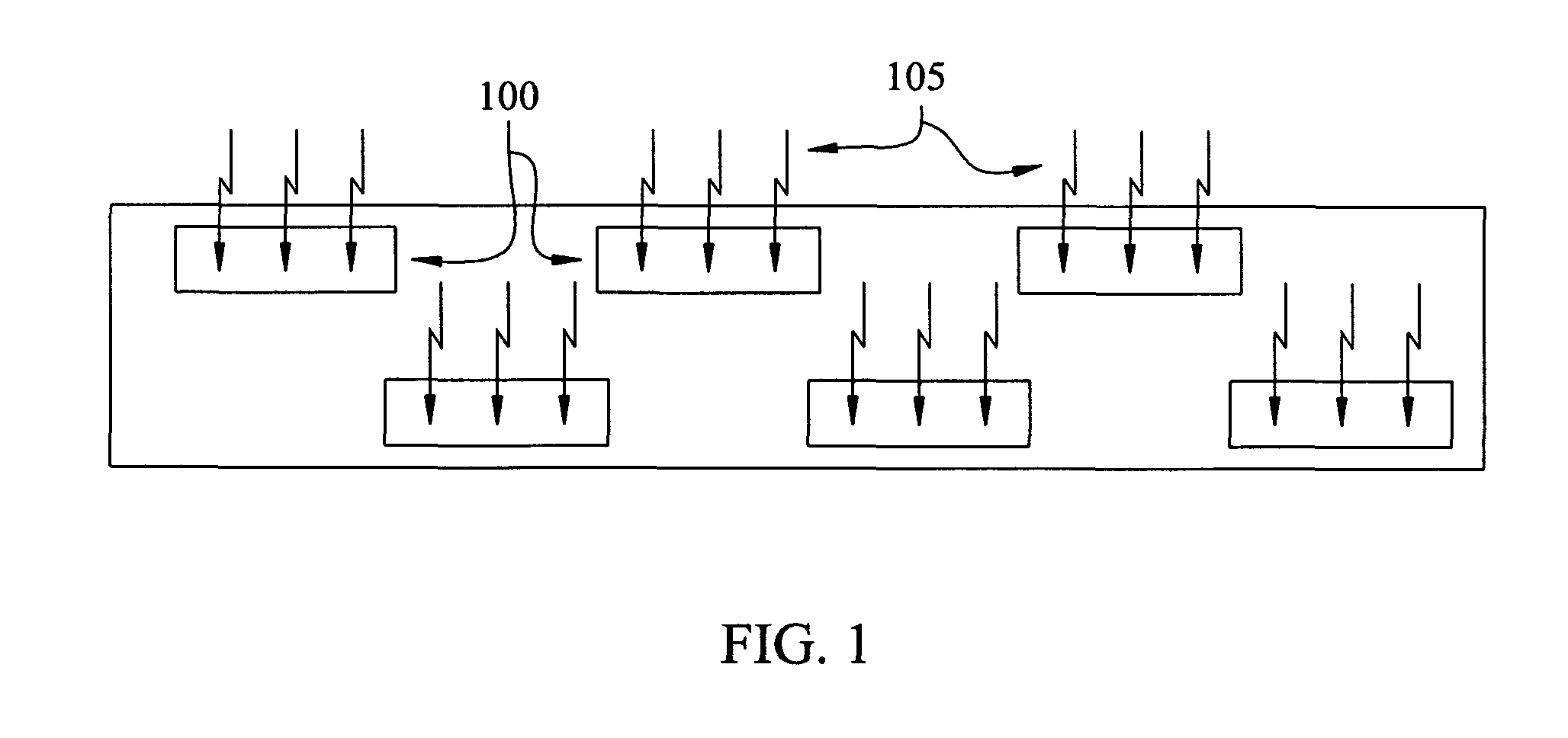
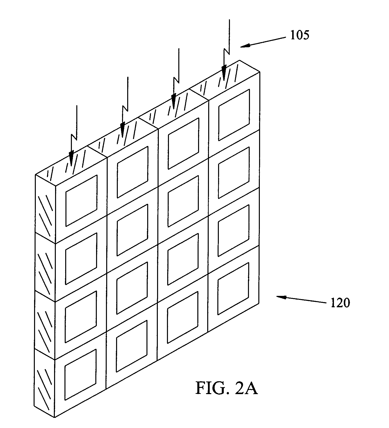
[0029]The invention provides edge-on and face-on scintillator detector designs and systems for enhanced slit and slot scan radiographic imaging for medical (including radiation therapy portal and CT, conventional CT), industrial, and scientific imaging applications. The scintillator-based detector for x-ray slit and slot scanning is comprised of: a scintillator x-ray detector, a photodetector coupled to the scintillator, high speed electronics for analyzing the readout signals using energy integration or photon counting techniques, and an electronic communications link to a computer for data post-processing, storage, and display. Additional aspects of the detector design include temperature control and power management as well as appropriate shielding from x-rays.

[0030]The general properties of edge-on and face-on detectors or detector modules (comprised of edge-on or face-on scintillator detectors, photodetector readout and processing electronics, focused and dynamic scanning capab...


PUM


Abstract

The invention provides methods and apparatus for detecting radiation including x-ray photon (including gamma ray photon) and particle radiation for radiographic imaging (including conventional CT and radiation therapy portal and CT), nuclear medicine, material composition analysis, container inspection, mine detection, remediation, high energy physics, and astronomy. This invention provides novel face-on, edge-on, edge-on sub-aperture resolution (SAR), and face-on SAR scintillator detectors, designs and systems for enhanced slit and slot scan radiographic imaging suitable for medical, industrial, Homeland Security, and scientific applications. Some of these detector designs are readily extended for use as area detectors, including cross-coupled arrays, gas detectors, and Compton gamma cameras. Energy integration, photon counting, and limited energy resolution readout capabilities are described. Continuous slit and slot designs as well as sub-slit and sub-slot geometries are described, permitting the use of modular detectors.

Description

[0001]This is a continuation of provisional application No. 61 / 123,238 filed on Apr. 8, 2008.FIELD OF THE INVENTION[0002]This invention provides novel face-on, edge-on, edge-on sub-aperture resolution (SAR), and face-on SAR scintillator and gas detectors, designs and systems for enhanced slit and slot scan area radiographic imaging and nuclear medicine imaging that are also suitable for industrial, Homeland Security, and scientific imaging applications.BACKGROUND OF THE INVENTION[0003]The dominant x-ray radiographic imaging system formats are the large area (flat) detectors and the slit / slot scanning detectors. Large area x-ray detectors are employed (in most cases) in stationary configurations in a face-on geometry. They tend to be expensive to build due to readout response uniformity problems over large areas. Response uniformity problems also limit the thickness of the detector material (typically a scintillator or semiconductor, although gas detectors have been used for applicat...

Claims


Application Information

Patent Timeline
Patent Type & Authority Patents(United States)
IPC IPC(8): G01D18/00
CPCG01T1/2018G01T1/202G01T1/2008G01T1/20183G01T1/20185G01T1/20181G01T1/20182
Inventor NELSON, ROBERT SIGURDNELSON, WILLIAM BERT
Owner MINNESOTA IMAGING & ENG
Why Eureka
Try Eureka