
RBCG (Bacillus Calmette-Guerin) expressing Brucella melitensis P39 gene as well as construction method and application of rBCG
A technique for species of Brucella and Brucella is applied in the field of rBCG expressing the P39 gene of Brucella species and its construction to achieve the effect of saving costs
Patent Information
- Authority / Receiving Office
- CN · China
- Patent Type
- Applications(China)
- Current Assignee / Owner
- Publication Date
- 2017-05-17
Smart Images
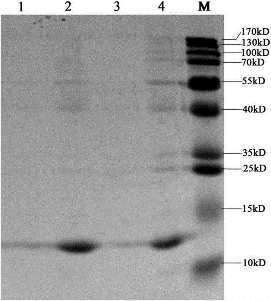
Figure 1 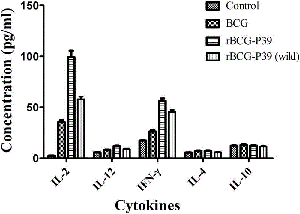
Figure 2
Abstract
Description
technical field
[0001] The invention relates to the technical field of genetic engineering and vaccine preparation, in particular to an rBCG expressing the P39 gene of Brucella melis and its construction method and application. Background technique
[0002] Brucellosis, referred to as brucellosis, is a zoonotic infectious disease caused by Brucella infection. Brucella is a facultative intracellular parasite that is highly infectious and pathogenic to humans and mammals. The genus mainly includes 6 species of sheep, cattle, pig and dog. The first 3 species are mainly prevalent in China, among which Brucella melis infection is the most common. The modes of transmission of brucellosis are animal-animal and animal-human. Livestock such as sheep, cattle, and pigs are most susceptible to infection. Infection of animals can lead to inflammation of reproductive organs and fetal membranes, miscarriage, infertility, and various tissue lesions, which greatly increases the chance of t...
Examples
Embodiment 1
[0030] Embodiment 1 carries the construction of the recombinant expression vector of Brucella melii P39 gene
[0031] Include the following steps:
[0032] 1. According to the published sequence of the P39 gene of Brucella melis M5 strain (GenBank: EF189139.1), after codon optimization using Jcat software, the P39 gene after the full sequence optimization was artificially synthesized as the target gene (nucleotide The sequence is shown in SEQ ID NO: 1).
[0033] 2. Construction of the recombinant expression vector carrying the optimized M5 strain P39 gene:
[0034] The above target gene was inserted into the shuttle expression vector pMV361 through two restriction sites of PvuⅡ and EcoRI.
[0035] 3. Verification of inserting the correct recombinant expression vector:
[0036] Through nucleic acid sequence determination, it was confirmed that the recombinant expression vector carrying the optimized Brucella melii P39 gene was constructed successfully.
Embodiment 2
[0037] The construction of embodiment 2 expressing the rBCG of Brucella melis P39 gene
[0038] 1. Using BCG as the host bacterium, transform the recombinant expression vector constructed in Example 1 (full sequence shown in SEQ ID NO: 2) into BCG.
[0039] The electroporation method was used for transformation, and the experimental conditions were: 2500V, 25μF, 1000Ω, electroporation time 5ms, 0.1cm electric shock cup. The electroporation reaction system is: 3 μl of plasmid (concentration is 0.65 μg / μl), 100 μl of competent BCG bacterial solution (concentration is about 1×10 10 CFU / ml).
[0040] 2. Screening of positive clones
[0041] After electroporation, the bacterial solution was drawn and inoculated on the medium (slant) containing 50 μg / ml kanamycin for positive clone selection.
[0042] 3. Detection of target gene expression
[0043] The screened positive clones (ie, recombinant BCG) were inoculated into liquid medium for expansion culture, the culture supernatant...
Embodiment 3
[0044] Effect experiment of embodiment 3 brucellosis vaccines
[0045] Immunize female Balb / c mice aged 6-8 weeks with recombinant BCG, inject subcutaneously, and the injection dose is 4×10 8 CFU / mouse, 4 weeks after immunization, the expression of Th1 / Th2 cytokines in the serum of mice in each group was detected. The experimental results showed that, compared with the recombinant BCG (rBCG-P39(wild)) carrying the unoptimized P39 gene and untransformed BCG, the recombinant BCG carrying the codon-optimized P39 gene (rBCG-P39) could effectively induce Production of Th1 cytokines such as IL-2, IL-12 and IFN-γ. ( figure 2 )