
Non-dividing donor cells for gene transfer
a donor cell and gene technology, applied in the field of non-dividing donor cells for gene transfer, can solve the problem of reducing the survival rate of reca mutants
Patent Information
- Authority / Receiving Office
- US · United States
- Patent Type
- Applications(United States)
- Current Assignee / Owner
- Publication Date
- 2006-01-05
- Estimated Expiration
- Not applicable · inactive patent
Smart Images
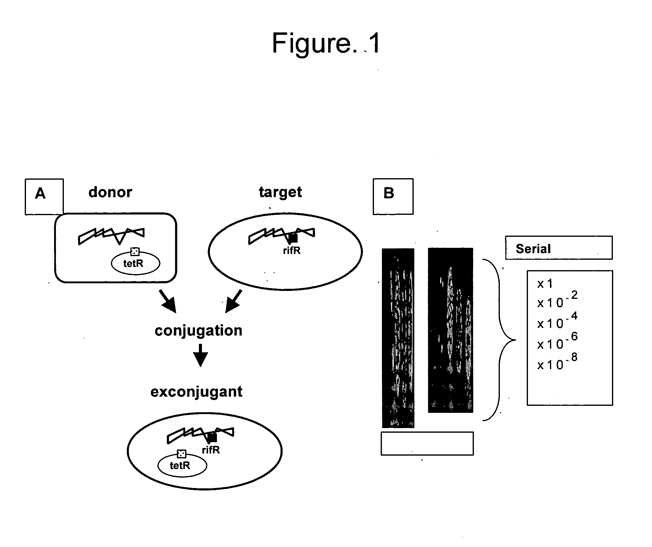
Figure 1 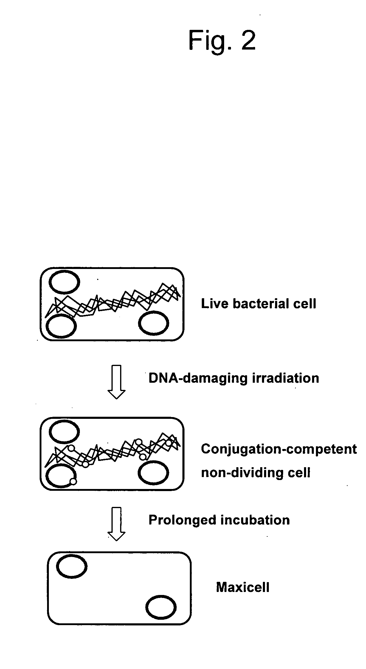
Figure 2 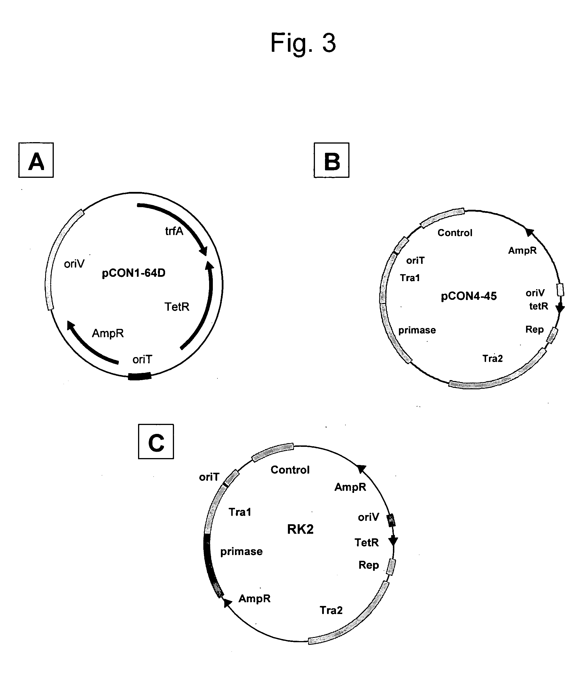
Figure 3
Abstract
Description
FIELD OF THE INVENTION
[0001] The present invention relates to compositions and methods for delivering genetic material to bacterial cells by conjugation with non-dividing donor cells. BACKGROUND OF THE INVENTION
[0002] Use of bacteria for numerous treatment purposes is well known in the art. For example, preparations of Lactobacillus acidophilus for use in human therapies is known (see, e.g., U.S. Pat. No. 5,032,399, Issued July, 1991 to Gorbach et al., and U.S. Pat. No. 5,733,568, issued March, 1998 to Ford). In addition, pharmaceutical preparations of Lactobacillus acidophilus are known (see., e.g., U.S. Pat. No. 4,314,995, issued Feb. 9, 1982 to Hata et al., “Pharmaceutical lactobacillus preparations”). Additional applications of bacteria in human therapeutics are described in U.S. Pat. No. 5,607,672 (Using recombinant Streptococcus mutans in the mouth to prevent tooth decay); U.S. Pat. No. 6,447,784 (Genetically modified tumor-targeted bacteria (Salmonella) with reduced virulen...
Examples
example 1
Plasmid Construction
[0116] This example describes the construction of exemplary plasmids to for use in the methods and compositions of the present invention.
A. Materials and Methods
Bacterial Strains and Media
[0117] The Escherichia coli strain utilized was JM109 [F′ traD36 proA+B+ laclq Δ (lacZ) M15 / Δ(lac-proAB) glnV44 el4− gyrA96 recA1 relA1 endA1 hsdR17]. All cloning was performed using standard methods known in the art, and using Luria Bertani growth media supplemented with 50 μg / ml kanamycin and / or 15 μg / ml tetracycline and / or 100 μg / ml ampicillin to permit selection for plasmids.
B. Plasmid Construction
Construction of pCON4-45
[0118] A DNA adaptor comprising multiple cloning sites (NsiI-MluI-NheI-SacI-AsiSI) was made by hybridizing a pair of DNA oligomers (5′ TACGCGTGCTAGCGAGCTCATTAATGCGAT 3′, SEQ ID NO:1, and 5′ CGCATTAATGAGCTCGCTAGCACGCGTATGCA3′, SEQ ID NO:2). The duplex adapter was cloned into the NsiI-AsiSI site of natural plasmid RK2 (diagrammed in FIG. 3C; Pansegr...
example 2
Bacterial Conjugation
[0120] This example provides an exemplary description of a method to perform bacterial conjugation.
A. Materials and Methods
Bacterial Strains and Media
[0121] The Escherichia coli strains utilized were S17-1 (Simon et al, Bio / Technology 1:784-791 [1985]), JM109 (Yanish-Perron et al., Gene 33:103-119 [1985]) and RL315. S17-1 is a K12-derived, non-pathogenic strain which is widely used in research laboratories working on bacterial conjugation. This strain carries the entire set of tra genes (derived from RK2) integrated into its chromosome, whose expression facilitates conjugal transfer of a resident mobilizable plasmid to a recipient. JM109 is also a K12-derived strain, and its relevant genotype is recA minus. S17-1 is also recA deficient, which harnesses the process of DNA-repair function after DNA-damaging irradiation. S17-1 was utilized to generate conjugation-competent non-dividing cells. Another K12-derived E. coli strain RL315 was utilized as a recipien...
example 3
Conjugal Transfer of a Self-Transmissible Plasmid from Non-Dividing JM109
[0123] This example demonstrates the transfer of a self-transmissible plasmid, RK2, from a non-dividing cell to a recipient cell, and shows that JM109 is a suitable strain for generating non-dividing cells using the method described in Example 3.
[0124]E. coli strain JM109 carrying the recA mutation was grown overnight in LB medium containing appropriate antibiotics. The cells were spun down, re-suspended in 0.9% NaCl and adjusted to OD600 1.0 prior to UV irradiation. Ten to fifteen milliliters of the cell suspension was transferred into a Petri dish, which was placed on a rotary shaker. A UV illuminator [302 nm] was placed inverted above the rotary shaker. The intensity of UV and the distance between the surface of the cell suspension and the UV lamp were kept constant. The cell suspensions were exposed to the UV light on the rotary shaker at 60 rpm, and bacterial cells were collected at different dosages of ...