
Compositions and Methods Relating to Treatment of Cancer and Infectious Diseases
a cancer and infectious disease technology, applied in the field of compositions and methods, can solve the problems of increasing tumour growth and ineffective killing of tumours, and achieve the effect of suppressing direct cell-to-cell contact and preventing suppression of the pro-inflammatory immune respons
Patent Information
- Authority / Receiving Office
- US · United States
- Patent Type
- Applications(United States)
- Current Assignee / Owner
- Publication Date
- 2010-01-07
- Estimated Expiration
- Not applicable · inactive patent
Smart Images
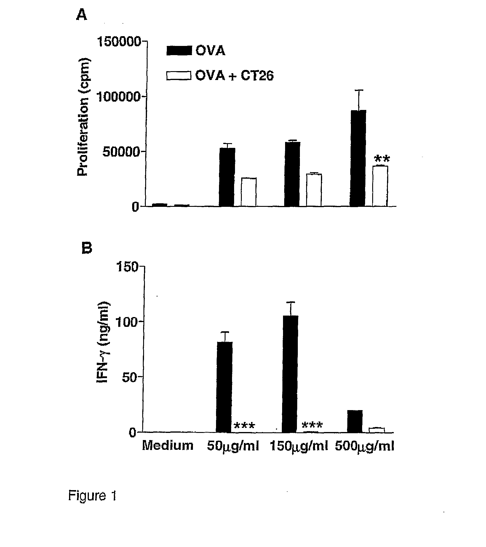
Figure 1 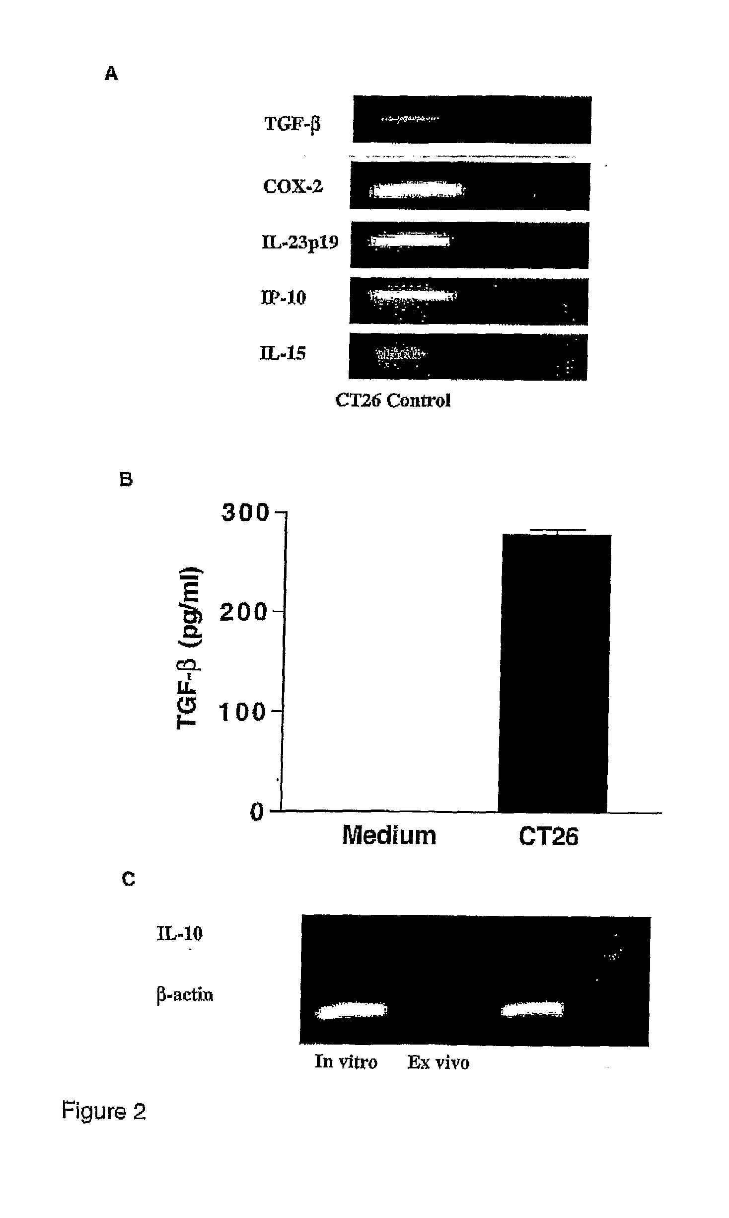
Figure 2 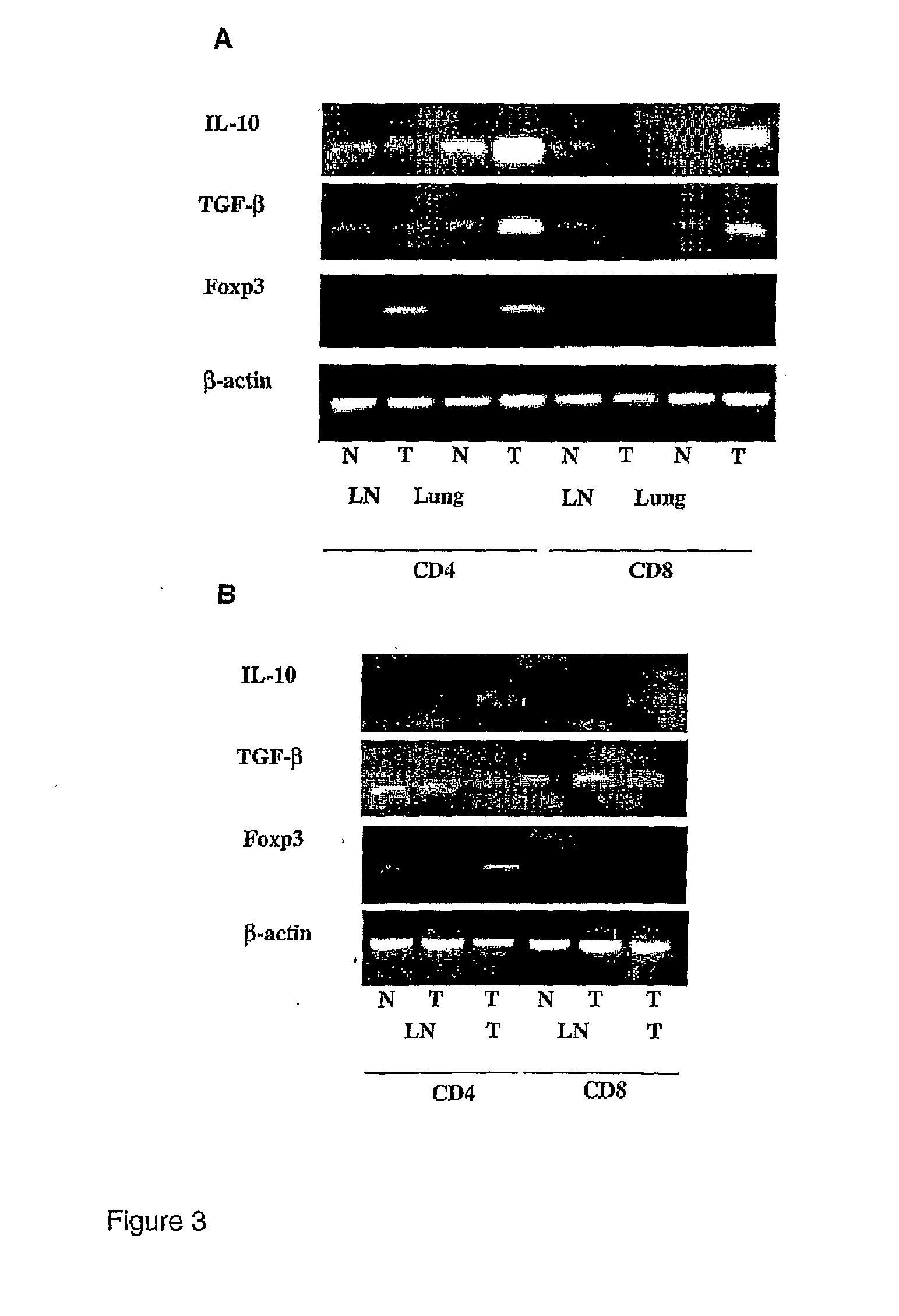
Figure 3
Abstract
Description
FIELD OF THE INVENTION
[0001] The present invention relates to novel compositions and methods for preventing or treating cancer or malignant conditions and infectious diseases. In particular, the invention relates to a composition and method for promoting the induction of type 1 immune responses (such as Th1 cells) and / or the subversion of regulatory T (Treg) cells. The invention further extends to uses of the compositions of the invention in the treatment of disease.BACKGROUND TO THE INVENTION
[0002] Cells of the innate immune system, especially dendritic cells (DCs), direct the differentiation of naïve CD4+ T cells into functionally distinct Th1, Th2 or regulatory T (Treg) cell subtypes. Activation of immature DCs through binding of conserved microbial molecules to pathogen recognition receptors (PRRs), such as Toll-like receptors (TLR) and integrins, is accompanied by maturation and homing to the lymph nodes, where the mature DCs present antigen (Ag) to the naïve T cells. Activatio...
Examples
example 1
CT26 Tumour Growth Inhibits T Cell Responses to Unrelated Antigens
[0287]Materials and Methods
[0288]DO.11.10 mice were injected s.c. with OVA (200 μg) alone or with 2×10⁵ CT26 cells. Mice were boosted after 7 days with 200 μg OVA and 14 days later lymph node cells were re-stimulated with OVA and proliferation (FIG. 1A) was examined after 4 days and IFN-gamma (FIG. 1B) concentrations determined in supernatants removed after 3 days. Results are mean±SD for 5 mice per group and assayed in triplicate. OVA versus OVA+CT26, **p<0.01, ***p<0.001 by ANOVA.
[0289]Results
[0290]In order to test the possibility that the failure to generate effective adaptive immune responses during tumour growth may result from an immunosuppressive environment created by the growing tumour, we examined the influence of a growing tumour on T cell responses to unrelated antigen. OVA-TCR Tg mice were injected s.c. with OVA in the presence or absence of CT26 cells and mice were boosted with OVA after 7 days, and sacr...
example 2
Immunosuppressive Cytokine Production Induced by Growing Tumours
[0291]Materials and Methods
[0292]RNA was extracted from cultured CT26 cells and RT-PCR was performed. using primers specific for IL-10, TGF-beta, IL-23p19, IL-15 and IP-10 (FIG. 2A). TGF-beta protein in supernatants of cultured CT26 cells was quantified by ELISA (FIG. 2B). RNA was extracted from in vitro cultured CT26 cells or homogenized solid CT26 tumours excised from mice bearing s.c. CT26 tumours (RNA pooled from 5 mice) and RT-PCR was performed using primers specific for IL-10 and beta-actin (FIG. 2C). Results are representative of 3 experiments.
[0293]BALB / c mice (5 per group) were injected with CT26 cells either i.v. (FIG. 3A) or s.c. (FIG. 3B). Lymph nodes (LN) and s.c. tumour masses (T) or lungs were taken from naïve (N) and tumour-bearing mice (T) 14 days after tumour challenge. CD4+ and CD8+ T cells were isolated using magnetic cell sorting and RNA was isolated and RT-PCR was performed using primers specific f...
example 3
Foxp3 Expression is Enhanced in CD4+ T Cells Within the CT26 Tumour Mass
[0297]Materials and Methods
[0298]BALB / c mice (5 per group) were injected with CT26 cells either intravenously (i.v.) (FIG. 3A) or subcutaneously (s.c.) (FIG. 3B). Lymph nodes (LN) and s.c. tumour masses (T) or lungs were taken from naïve (N) and tumour-bearing mice (T) 14 days after tumour challenge. CD4+ and CD8+ T cells were isolated using magnetic cell sorting and RNA was isolated and RT-PCR was performed using primers specific for Foxp3.
[0299]Results
[0300]Having demonstrated that T cells expressing IL-10 and TGF-beta accumulate in the tumour during growth, we examined the possibility that natural Treg cells were recruited to the site of the tumour. CD4+ and CD8+ T cells were purified from inguinal lymph nodes and solid tumours in the s.c. model and superficial lymph nodes and lungs in the lung metastasis model and RT-PCR was performed using primers specific for Foxp3. In the lung metastasis model, Foxp3 expr...