Method for segmenting renal cortex images

An image segmentation and renal cortex technology, applied in the field of image processing, can solve the problems of renal cortex image failure, difficult renal cortical structure segmentation, etc., and achieve the effects of inhibiting renal cortex from embedding renal columns, reducing workload, and overcoming image noise.

Inactive Publication Date: 2012-01-25
INST OF AUTOMATION CHINESE ACAD OF SCI
View PDF3 Cites 6 Cited by
  • Summary
  • Abstract
  • Description
  • Claims
  • Application Information

AI Technical Summary

Problems solved by technology

[0005] In order to solve the problem that the existing technology is difficult to accurately segment the renal cortex structure, which leads to the failure of conventional methods in

Method used


Image

  • Method for segmenting renal cortex images
  • Method for segmenting renal cortex images
  • Method for segmenting renal cortex images

Examples


Embodiment Construction

[0026] In order to make the object, technical solution and advantages of the present invention clearer, the present invention will be described in further detail below in conjunction with specific embodiments and with reference to the accompanying drawings.
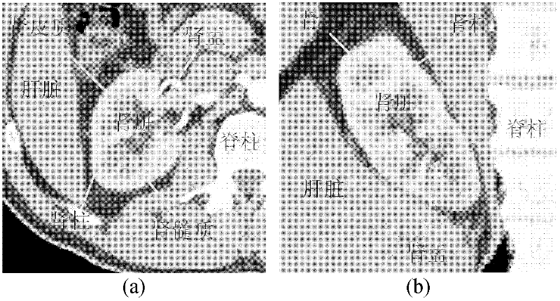
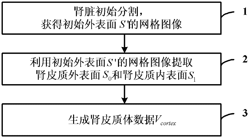
[0027] The core idea of ​​the present invention is to propose a renal cortex image segmentation method to accurately segment the renal cortex structure. The method for segmenting renal cortex images provided by the present invention will be described in detail below in conjunction with specific embodiments, as figure 2 Shown is the flowchart of the kidney cortex image segmentation method provided by the present invention, and the method comprises the following steps:

[0028] Step 1: Use the statistical shape model algorithm to initially segment the kidney image to obtain the grid image of the initial outer surface S' of the kidney structure;

[0029] Step 2: In the narrow band near the grid image of the initial outer s...


PUM


Abstract

The invention discloses a method for segmenting renal cortex images. The method comprises the following steps of: 1, performing initial segmentation on a kidney image by using a statistical shape model algorithm to acquire grid images of an initial outer surface S' of a kidney structure; 2, establishing sub-graphs G0 and G1 for the outer surface and the inner surface of a renal cortex respectively in a narrow band close to the grid images of the initial outer surface S' so as to obtain a weighted directed graph G=(G0, G1), designing cost functions C0 and C1 by aiming at the outer surface and the inner surface of the renal cortex respectively, generating an s-t graph Gst based on the weighted directed graph G=(G0, G1) and the cost functions C0 and C1, and extracting two layers of optimal surface images, namely an outer surface image S0 and an inner surface image S1 of the renal cortex, by using a max-flow minimal cut method; and 3, generating a data image Vcortex of a renal cortex body by using the outer surface image S0 and the inner surface image S1 of the renal cortex. By the method, the renal cortex structure can be segmented accurately, and the workload for manually segmenting the renal cortex is greatly reduced, so the method has important application values in the field of segmenting the renal cortex images and ventricular wall images with multi-surface structural characteristics.

Description

technical field [0001] The invention relates to image processing technology, in particular to a renal cortex image segmentation method. Background technique [0002] With the development of imaging technology, medical image segmentation has become a key and challenging problem in the field of medical image analysis. Kidney segmentation is particularly complex based on the special structure of the kidney. Such as figure 1 Shown is a CT image of the kidney, which mainly consists of the renal cortex, renal medulla, and renal pelvis. The renal cortex forms a continuous smooth outer band, interspersed with a series of projections from the renal pelvis to the renal cortex, which are the renal columns. There are already some algorithms for kidney segmentation, but most of them focus on whole kidney segmentation and rarely involve specific structures in the kidney such as renal cortex and renal column. figure 1 (a) in represents the coronal plane; figure 1 (b) in (b) represents...

Claims


Application Information

Patent Timeline
IPC IPC(8): G06T7/00
Inventor 田捷李秀丽张星杨鑫
Owner INST OF AUTOMATION CHINESE ACAD OF SCI
Why Eureka
Try Eureka