3D intraoral scanner measuring fluorescence

a scanner and fluorescence technology, applied in the field of intraoral 3d scanning, can solve the problems of deteriorating the quality of the stitching algorithm result, and affecting the quality of the stitching algorithm. , the effect of improving the stitching quality

Active Publication Date: 2019-03-26
3SHAPE AS
View PDF25 Cites 5 Cited by
  • Summary
  • Abstract
  • Description
  • Claims
  • Application Information

AI Technical Summary

Benefits of technology

[0018]The mapped representation of an identified cariogenic regions may provide an improved visibility in relation to the tooth surface compared to how visible the identified cariogenic regions are on the tooth, such that the visualization of the combined digital 3D representation can provide valuable assistance to the dentist when examining a patient's set of teeth. This may e.g. be the case when the cariogenic region is represented with a distinct color and / or brightness in the combined digital 3D representation.
[0216]In some embodiments, the 3D scanner system is capable of adjusting the settings relating to the precision and / or spatial resolution at which the 3D scanner system acquires data from surface for computing the sub-scans, such that a higher precision and / or resolution can provided for the region of the digital 3D representation corresponding to the region of particular interest.

Problems solved by technology

The size of any probe element in intraoral scanners is limited because it has to fit into the human mouth.
In practice, the assumption of no relative movement of 3D scanner and intraoral cavity during the 3D surface and fluorescence recordings will generally not hold perfectly.
A particular problem with stitching of intraoral sub-scans is soft tissue in the oral cavity that moves between sub-scans and even within a sub-scan.
Overlapping regions in multiple sub-scans may therefore not be identified correctly, deteriorating the quality of the stitching algorithm's result.
This results in the soft tissue wrapping around the sub-gingival preparation line and hinders scanning.

Method used


Image

  • 3D intraoral scanner measuring fluorescence
  • 3D intraoral scanner measuring fluorescence
  • 3D intraoral scanner measuring fluorescence

Examples


Embodiment Construction

[0285]In the following description, reference is made to the accompanying figures, which show by way of illustration how the invention may be practiced.

[0286]The figures below illustrate schematically how embodiments of a 3D scanner according to this invention can be realized. The figures are not necessarily dimensionally precise.

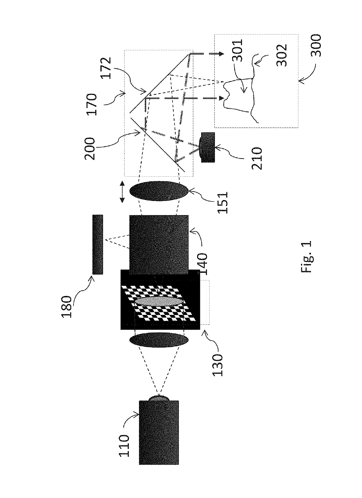
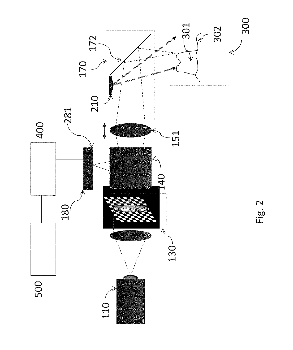
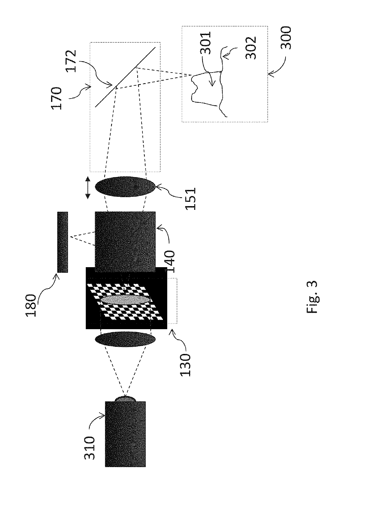
[0287]FIG. 1 shows an embodiment of the invention, namely a focus scanning intraoral 3D scanner comprising an illumination unit with a first light source 210 and a second light source 110, a pattern 130 (a line in a true cross-sectional view, but shown here at an angle for clarity), an image sensor 180, a beam splitter 140, and focusing optics with a moveable lens 151. The 3D scanner has a tip or probe 170 with a mirror 172 that folds the beam path towards the region of the intraoral cavity being scanned 300. The intraoral cavity comprises hard dental tissue 301 and soft dental tissue 302. The second light source 110 emits light at least at the second wavel...


PUM


Abstract

Disclosed is a 3D scanner system for detecting and / or visualizing cariogenic regions in teeth based on fluorescence emitted from the teeth, said 3D scanner system including a data processor configured for mapping a representation of fluorescence emitted from the teeth onto the corresponding portion of a digital 3D representation of the teeth to provide a combined digital 3D representation.

Description

FIELD OF THE INVENTION[0001]The invention relates to intraoral 3D scanning. In particular the invention relates to a 3D scanner system where fluorescence recorded from a patient's teeth is used for detecting cariogenic regions on the teeth. In particular the invention relates to a 3D scanner system where fluorescence recorded from the intraoral cavity is used when generating a digital 3D representation of the intra oral cavity.BACKGROUND OF THE INVENTION[0002]US20080063998 describes a technique wherein one fluorescence image and one reflectance image are combined to form an image in which the contrast between a cariogenic region and a sound tooth structure is heightened in the 2D image.[0003]US2008094631 describes a system with intraoral camera and measuring device. The measuring device is capable of collecting color, translucency, fluorescence, gloss, surface texture and / or other data for a particular tooth from the measuring device which then may be combined with images captured b...

Claims


Application Information

Patent Timeline
Patent Type & Authority Patents(United States)
IPC IPC(8): A61C3/00A61B5/00A61C9/00
CPCA61B5/0088A61B5/0071A61C9/0053A61B5/742A61B5/0035A61B5/0082
Inventor VAN DER POEL, MIKEHOLLENBECK, KARL-JOSEF
Owner 3SHAPE AS
Why Eureka
Try Eureka