
Endometrial Phase or Endometrial Cancer Biomarkers
a biomarker and endometrial technology, applied in the field of endometrial markers, can solve the problems of inability to screen, inability to apply, and inability to achieve the effect of screening
- Summary
- Abstract
- Description
- Claims
- Application Information
AI Technical Summary
Benefits of technology
Problems solved by technology
Method used
Image

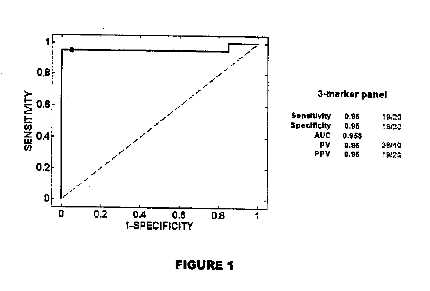
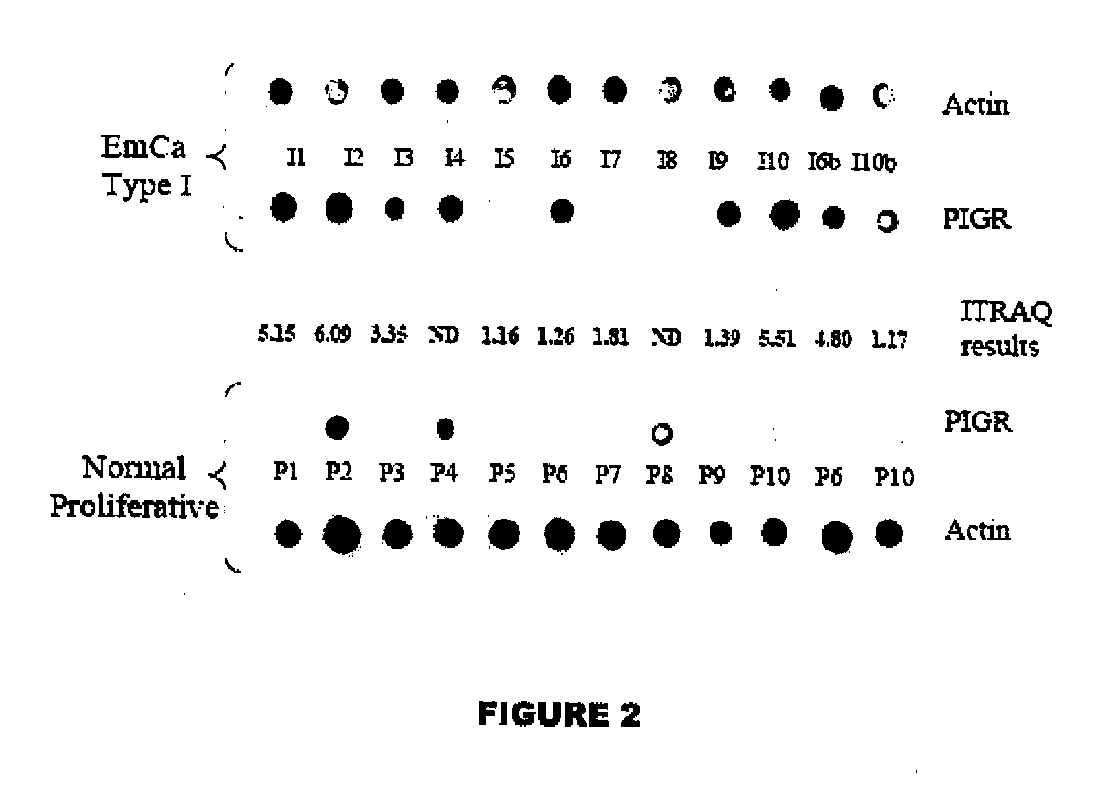
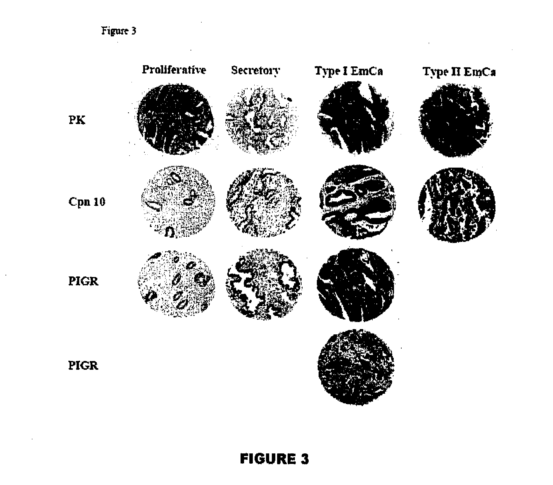
Examples
example 1
Experimental Procedures
Samples and Reagents
[0401]Endometrial tissues were retrieved from an in-house, dedicated, research endometrial-tissue bank. With patient consent, samples from hysterectomy specimens had been flash-frozen in liquid nitrogen within 20 minutes of devitalizing. The patient consent forms and tissue-banking procedures were approved by the Research Ethics Boards of York University, Mount Sinai Hospital, University Health Network, and North York General Hospital. These frozen samples were sectioned and stored at −80° C. The histologic diagnosis for each sample was confirmed using microscopic examination of a hematoxylin and eosin-stained frozen section of each research tissue block. The tissue from the mirror face of the histologic section was then washed three times in approximately 1 mL of phosphate-buffered saline (PBS) with a cocktail of protease inhibitors as described previously (1 mM AEBSF, 10 μM leupeptin, 1 μg / mL aprotinin, and 1 μM pepstatin) (3). The washed...
PUM
 Abstract
Description
Claims
Application Information