Combined PET/MR imaging system and APD-based pet detector for use in simultaneous PET/MR imaging

a combined imaging system and detector technology, applied in the field of medical imaging and systems for obtaining diagnostic images, can solve problems such as complex design requirements and inability to achieve high performance characteristics

Active Publication Date: 2015-09-01
SIEMENS MEDICAL SOLUTIONS USA INC
View PDF24 Cites 8 Cited by
  • Summary
  • Abstract
  • Description
  • Claims
  • Application Information

AI Technical Summary

Problems solved by technology

However, such designs have not been able to achieve high performance characteristics and suffer from complicated design requirements.

Method used


Image

  • Combined PET/MR imaging system and APD-based pet detector for use in simultaneous PET/MR imaging
  • Combined PET/MR imaging system and APD-based pet detector for use in simultaneous PET/MR imaging
  • Combined PET/MR imaging system and APD-based pet detector for use in simultaneous PET/MR imaging

Examples


example implementation

[0025 and Test Measurement

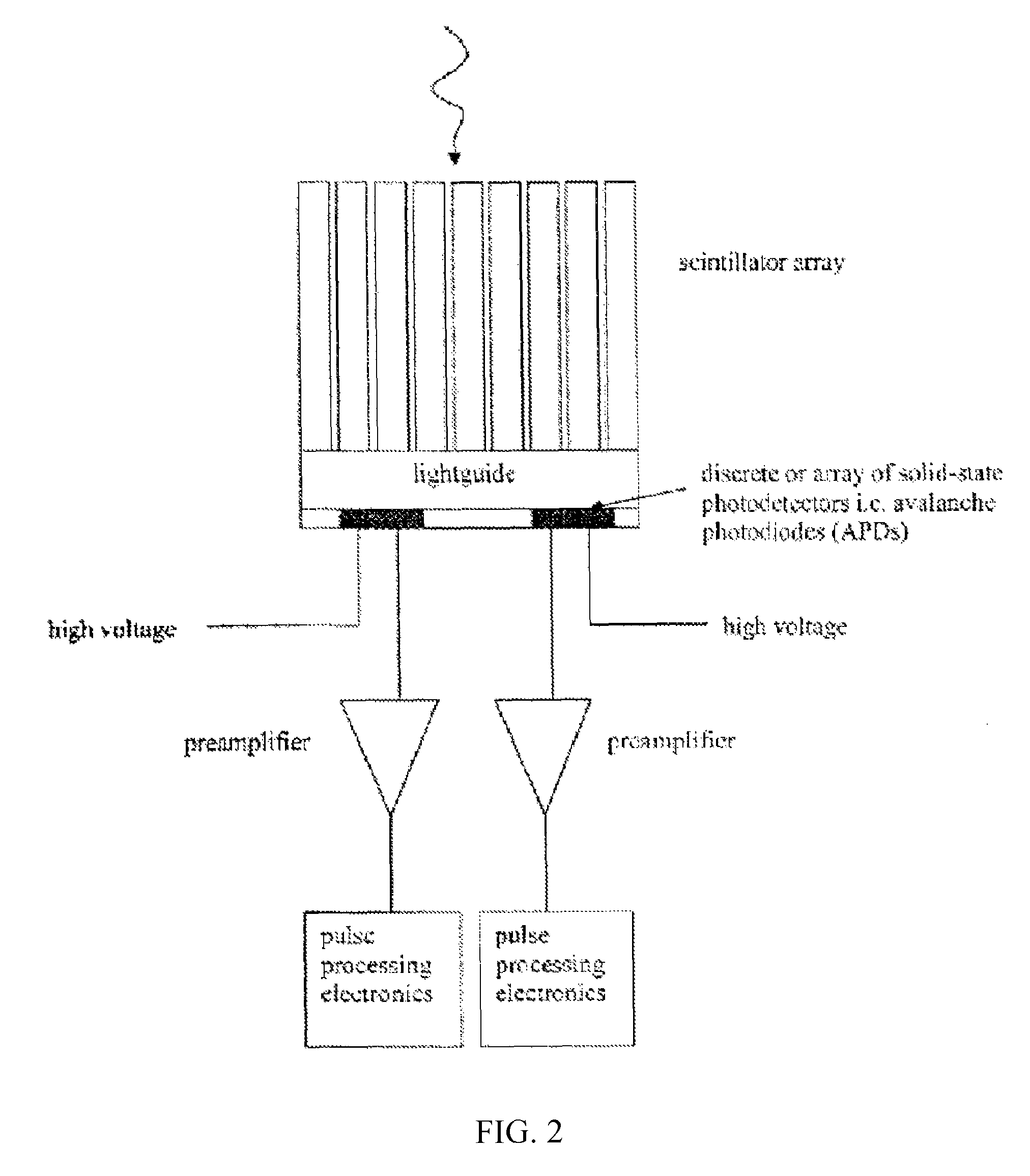
[0026]An APD-based PET module has been built and tested for use in a MR scanner for simultaneous PET / MR imaging according to the present invention. The module consisted of 4 optically isolated scintillator blocks each read out by a 2×2 APD array, as shown in FIG. 5. One basic APD detector design according to an embodiment of the invention is based on an LSO block design. The scintillator blocks are 8×8 arrays of 2 mm×2 mm×20 mm LSO crystals coupled to glass light guides. The APDs are coupled to the glass light guides. The APDs can be any commercially available APD, such as, e.g., Hamamatsu S8664-55 APDs in a custom package, or APDs available from Perkin-Elmer. The APD signals are amplified by a charge-sensitive preamplifier ASIC, and shaped by a pole-zero circuit. Therefore one module contained 4 LSO blocks, 16 APDs, 2 ASIC preamplifiers, and 16 channels of pole-zero electronics.

[0027]The outputs of the modules were sampled and digitized by Siemens Pico3D e...


PUM


Abstract

APD-based PET modules are provided for use in combined PET / MR imaging. Each module includes a number of independent, optically isolated detectors. Each detector includes an array of scintillator (e.g. LSO) crystals read out by an array of APDs. The modules are positioned in the tunnel of a MR scanner. Simultaneous, artifact-free images can be acquired with the APD-based PET and MR system resulting in a high-resolution and cost-effective integrated PET / MR system.

Description

CLAIM OF PRIORITY FROM RELATED APPLICATIONS[0001]This application is a continuation of application Ser. No. 11 / 319,795 filed Dec. 29, 2005, which claims priority under 35 U.S.C. §119(e) from provisional applications Ser. No. 60 / 640,073 filed Dec. 29, 2004, and Ser. No. 60 / 738,998 filed Nov. 23, 2005.FIELD OF THE INVENTION[0002]The present invention generally relates to the field of medical imaging, and systems for obtaining diagnostic images such as nuclear medicine images and magnetic resonance (MR) images. In particular, the present invention relates to multiple modality imaging systems and methods for obtaining diagnostic images of multiple modalities, such as nuclear medicine images from positron emission tomography (PET) data and magnetic resonance imaging (MRI) data. The PET images and MR images can be obtained either sequentially or simultaneously.BACKGROUND OF THE INVENTION[0003]Nuclear medicine is a unique medical specialty wherein radiation is used to acquire images which ...

Claims


Application Information

Patent Timeline
Patent Type & Authority Patents(United States)
IPC IPC(8): G01T1/164A61B6/03G01T1/29G01T1/16G01R33/48G01R33/381G01R33/26G01R33/28G01T1/166A61B8/08
CPCG01R33/28A61B6/037G01R33/26G01R33/381G01R33/481G01T1/1603G01T1/2985A61B8/5261G01T1/20185G01T1/20182G01T1/20188A61B5/05A61B5/055G01R33/20G01V3/00G01R33/307
Inventor SCHMAND, MATTHIAS J.GRAZIOSO, RONNUTT, RONALDNUTT, ROBERT E.ZHANG, NANCORBEIL, JAMES LUKELADEBECK, RALFVESTER, MARKUSSCHNUR, GUNTERRENZ, WOLFGANGFISCHER, HUBERTUSPICHLER, BERND J.
Owner SIEMENS MEDICAL SOLUTIONS USA INC
Why Eureka
Try Eureka