
Test strip for detecting alpha fetoprotein in whole blood quickly and manufacturing method of test strip
A technology of alpha-fetoprotein and test strips, which is applied in the field of rapid detection of alpha-fetoprotein in whole blood test strips and its preparation, which can solve the problems of cumbersome operation steps of detection instruments, difficulties in popularizing households of ordinary citizens, and detection of serum enlargement Difficulty and other issues, to achieve the effect of high sensitivity, low detection cost, and reasonable structural design
Patent Information
- Authority / Receiving Office
- CN · China
- Current Assignee / Owner
- Publication Date
- 2015-12-16
- Estimated Expiration
- Not applicable · inactive patent
Smart Images
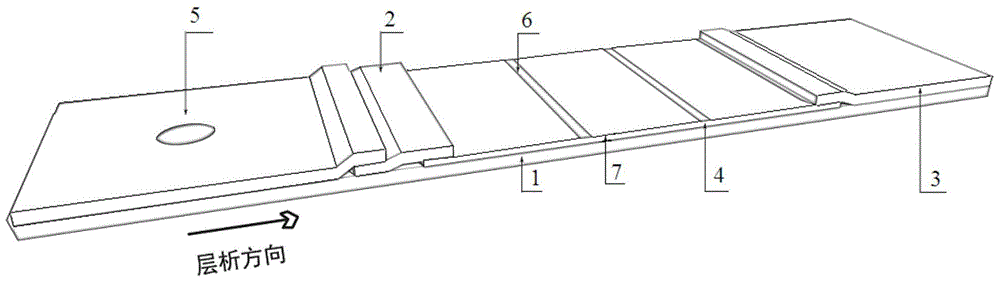
Figure 1 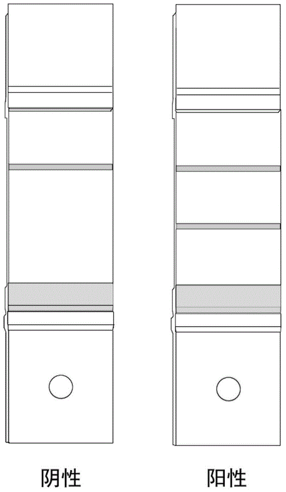
Figure 2 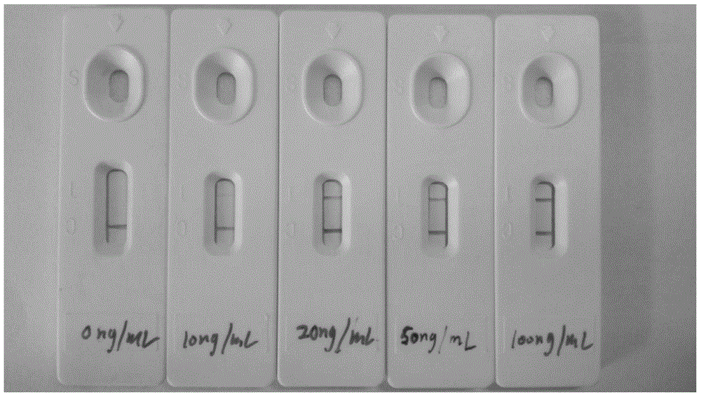
Figure 3
Abstract
Description
technical field
[0001] The invention belongs to the field of biological test product manufacture, and in particular relates to a test strip for rapidly detecting alpha-fetoprotein in whole blood and a preparation method thereof. Background technique
[0002] The rapid and accurate detection of liver cancer specific markers is of great significance for the early diagnosis and treatment of primary liver cancer. Under normal circumstances, alpha-fetoprotein (AFP) mainly comes from the liver cells of the embryo. About two weeks after the fetus is born, alpha-fetoprotein disappears from the blood. The serum alpha-fetoprotein of normal people is still less than 20ng / ml. During the period of liver cell regeneration, some embryonic carcinomas, germ cell tumors, digestive tract cancers, and AFP in the blood of pregnant women also increase to varying degrees. But the most noteworthy is that in patients with liver cancer, the concentration of AFP in the blood can be greatly increased,...
Examples
preparation example Construction
[0036] The preparation method of the test strip for rapid detection of alpha-fetoprotein in whole blood disclosed by the present invention comprises the following steps:
[0037] 1) Colloidal gold particles with uniform shape and size were prepared by the citrate reduction method of chloroauric acid, and the diameter of the particles was 20 nm.
[0038] 2) Prepare the colloidal gold immunoprobe, adjust its pH to 8.5-9.0, and modify the AFP monoclonal antibody 2 according to the principle that high-concentration salt solution can cause coagulation of unstable colloidal gold.
[0039] When modifying, mix 0.6-1.5ml of colloidal gold and 10-15μg of AFP monoclonal antibody 2 evenly and let stand at 4°C for 25-35 minutes. Add BSA for blocking for 35-45 minutes, keeping its final concentration at 0.5%-1.5%. Centrifuge at 10000-14000rpm under centrifugation conditions, and use PBS buffer containing 0.5%-1.5% BSA, 0.03%-0.1% sodium azide, 0.5%-1.5% sucrose, and 0.5%-1.5% Tween-20 for ...
Embodiment 1
[0044] 1) Preparation of 20nm colloidal gold: Take 100 mL of 0.01% (v / v) chloroauric acid solution, put it into a cleaned and dried Erlenmeyer flask, place it on a heating magnetic stirrer and heat it to boiling. Maintain the heating state and accurately and quickly add 2.0 ml of freshly prepared 1% (v / v) trisodium citrate solution in one go under vigorous stirring. It can be observed that the color of the solution changes from yellow→black→purple→dark blue→wine red, and continue stirring for 15 minutes until the solution turns transparent wine red. After the reaction solution was cooled to room temperature, it was stored at 4°C.
[0045] 2) Preparation of immune probe: first use 0.1M k 2 CO 3 The pH of the colloidal gold was adjusted to 8.7, and 13 μg of AFP monoclonal antibody 2 was added to 1 ml of the colloidal gold solution and left to stand at 4° C. for 25 minutes. 10% (v / v) BSA was added for blocking for 40 minutes, keeping its final concentration at 1%. Make the im...