
Larimichthys crocea testis tissue cell line and application thereof
A technology of histiocytes and large yellow croaker, applied in the biological field, can solve the problems that limit the research on the mechanism of gonad development of large yellow croaker, and achieve the effects of improving mitotic activity, inhibiting bacterial growth, and good stability
Patent Information
- Authority / Receiving Office
- CN · China
- Current Assignee / Owner
- Publication Date
- 2021-05-28
Smart Images
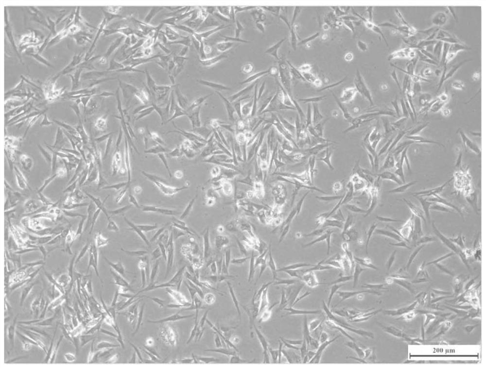
Figure 1 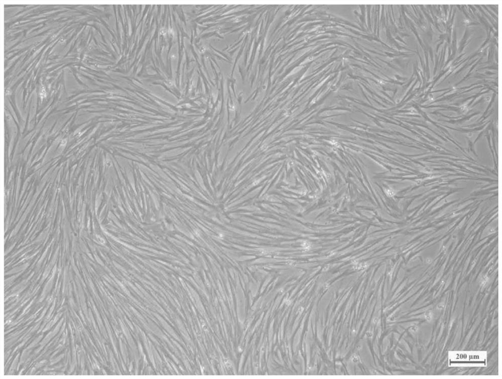
Figure 2 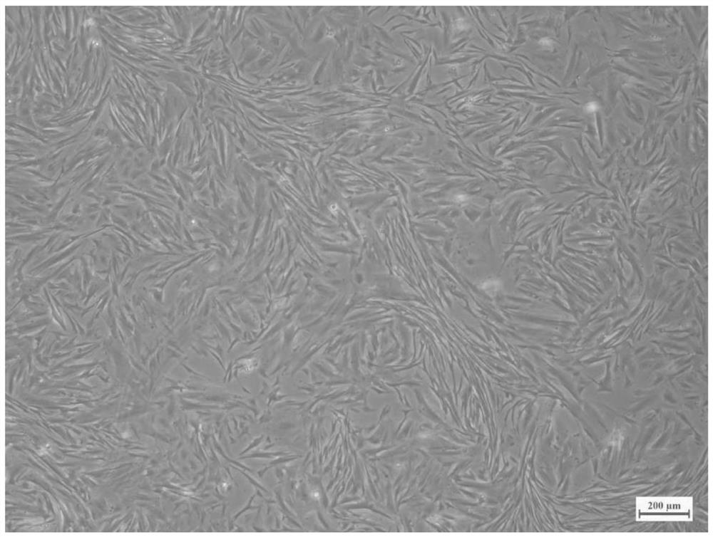
Figure 3
Abstract
Description
technical field
[0001] The invention relates to the field of biotechnology, in particular to a large yellow croaker testis tissue cell line and application thereof. Background technique
[0002] large yellow croaker ( Larimichthys crocea ), Osteostichthyes, Perciformes, Totoaceae, and Yellow Croaker, mainly inhabit the middle and lower layers of coastal and offshore waters, are the main economic fish in China's coastal waters, and are traditional Chinese "four seafood" (large yellow croaker, small yellow croaker , hairtail, squid). At present, the main production areas of large yellow croaker are Fujian, Zhejiang and Guangdong. In 2019, Fujian produced 186,514 tons, accounting for 82.7%; Zhejiang produced 23,932 tons, accounting for 10.6%; Guangdong produced 15,103 tons, accounting for 6.7%. However, factors such as the depletion of large yellow croaker broodstock resources, germplasm degradation, and frequent occurrence of large yellow croaker diseases have greatly restr...
Examples
Embodiment 1
[0054] Example 1 Establishment of testicular tissue cell line of large yellow croaker
[0055] S1. Obtaining the testis tissue of large yellow croaker: Since large yellow croaker is a fish of the Totoaba family and is extremely sensitive to hypochlorite, sterilized seawater containing double antibodies was used instead of hypochlorous acid disinfectant in the body surface disinfection step. The 7-month-old large yellow croaker fry with a length of 9-11 cm were temporarily raised in sterilized seawater containing double antibodies for 1-2 hours. At the end, 3 - 4 drops of eugenol were dripped until the fish turned over, the belly was up, and there was no stress response to the stimulation. Wipe off the mucus on the surface of the fish with a sterilized gauze piece. Wipe the surface of the fish twice with gauze soaked in 75% alcohol. Move the fish body into the ultra-clean bench, take out the testis tissue with dissecting instruments, rinse in PBS solution containing penicilli...
Embodiment 2
[0059] Example 2 Cryopreservation and recovery of large yellow croaker testis tissue cell line
[0060] S1. Freezing: Take a bottle of 75cm 2 The vigorous growth of large yellow croaker testis cell line cells covering the bottom of the culture bottle was digested with trypsin, and the cell pellet was collected after centrifugation, and 3 mL of the prepared cell freezing solution was slowly added, and the cells were blown gently to make them Evenly disperse in the cell freezing solution, and use a pipette to transfer the liquid into the cryovial. Place the cryopreservation tube at 4°C for 1 hour, then place it in a programmed gradient cooling freezer box (temperature drop rate is 1°C / min), place the freezer box at -80°C for 1 day, and finally take out the cryopreservation tube and dry it. Immersed in liquid nitrogen, can be frozen for a long time.
[0061] S2. Take the cryopreservation tube out of the liquid nitrogen and quickly place it in a water bath with adjusted temperat...
Embodiment 3
[0063] Example 3 Proliferation curves of different passage ratios of large yellow croaker testis tissue cell lines
[0064] From the large yellow croaker testis tissue cell line established in Example 1, cells with good morphology and vigorous proliferation were selected, digested and passaged, and passaged to 25 cm at 1:2, 1:3, 1:5 and 1:10, respectively. 2 Cell culture flasks (i.e. add 2.5 mL, 1.6 mL, 1 mL, 0.5 mL of cell suspension to the 4 culture flasks, and then make up the total amount of each bottle to 5 mL with culture medium). Photographs were taken every 24 hours from the 24th hour of subculture, and the cultured cells were taken by inverted microscope using the classic five-point cross sampling method to count the number of cells observed in the visual field, and the sum of the number of cells observed at five points in each group was plotted for analysis.
[0065] like Figure 14 It was shown that for the large yellow croaker testis tissue cell lines, different s...