

Detection of nucleic acid differences using endonuclease cleavage/ligase resealing reactions and capillary electrophoresis or microarrays
a nucleic acid and endonuclease technology, applied in microorganism testing/measurement, biochemistry apparatus and processes, nanoinformatics, etc., can solve the problems of patients facing the real possibility, unable to calibrate therapy to the molecular state of the tumor, and difficult to meaningfully predict individual treatment and stratification. , to achieve the effect of extending hairpins and reducing nois
Patent Information
- Authority / Receiving Office
- US · United States
- Patent Type
- Applications(United States)
- Current Assignee / Owner
- Publication Date
- 2009-05-14
- Estimated Expiration
- Not applicable · inactive patent
Smart Images
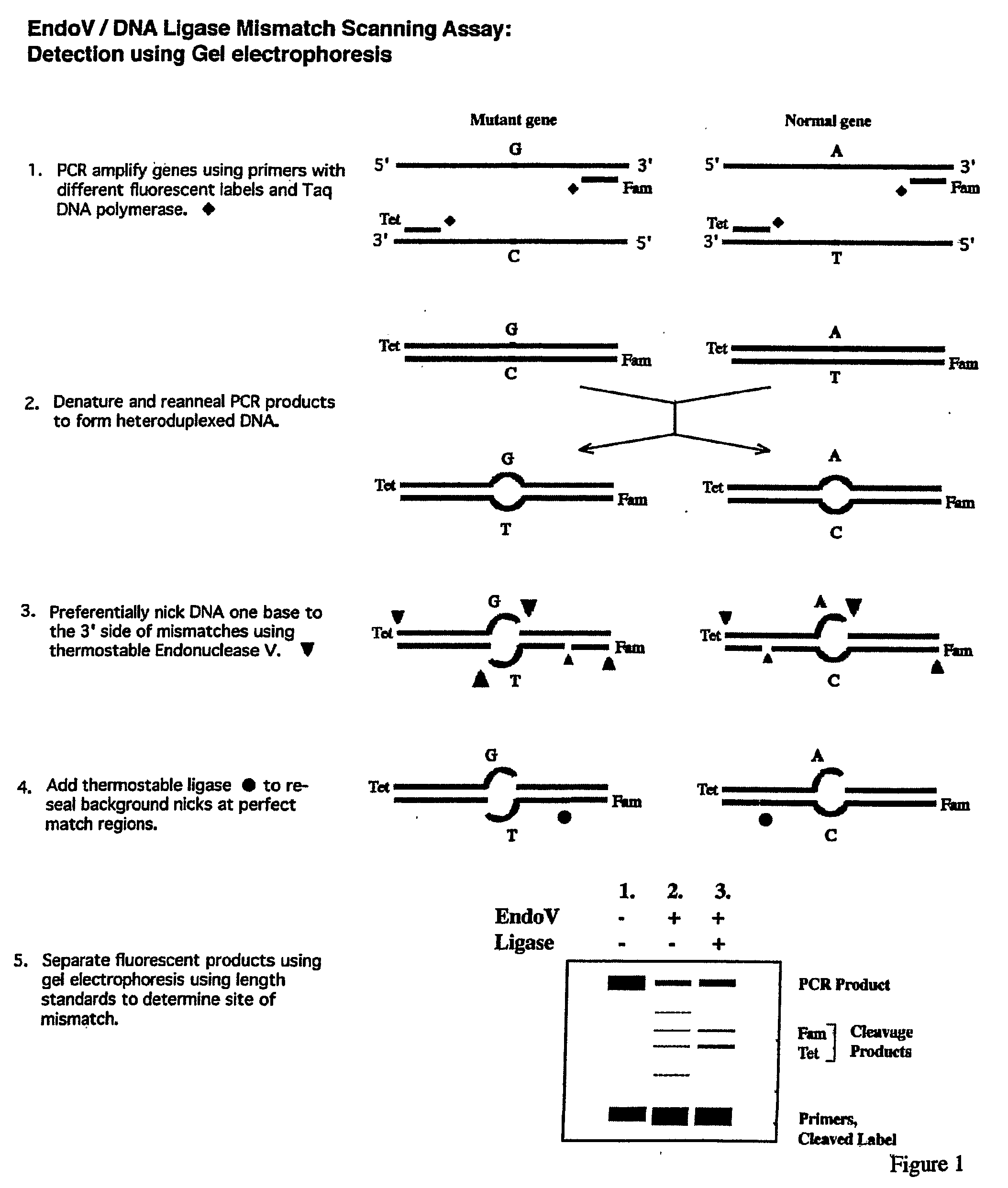
Figure 1 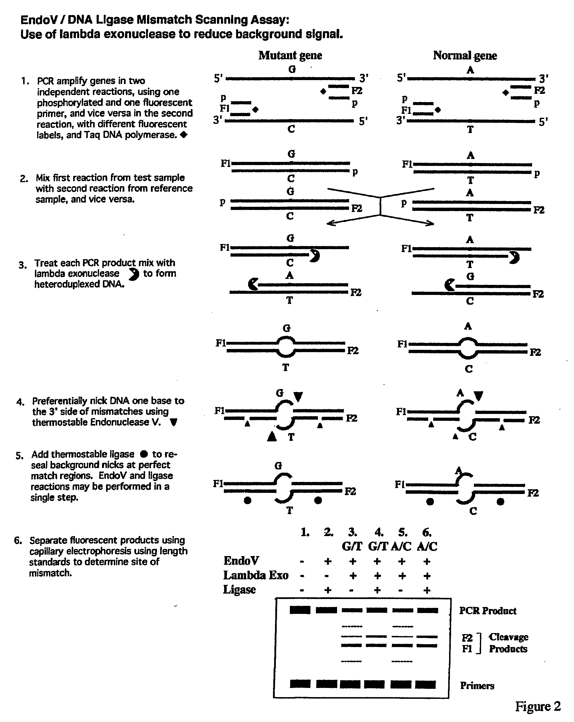
Figure 2 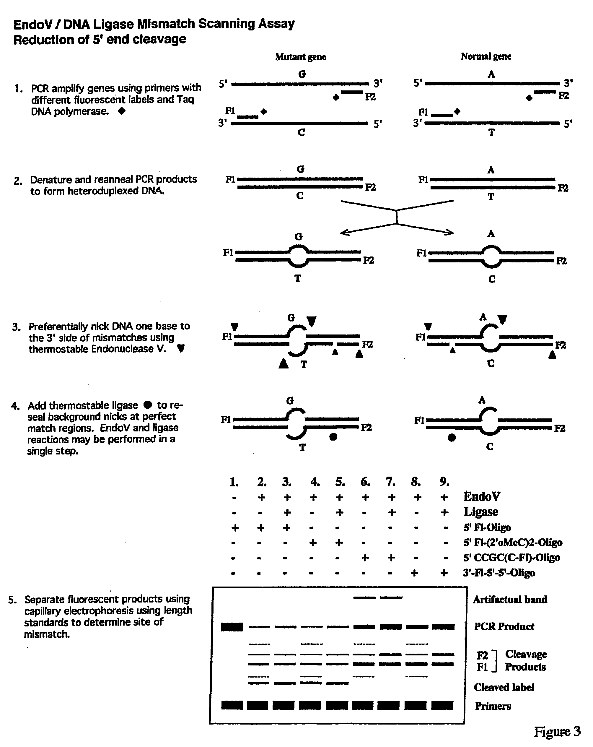
Figure 3
Abstract
Description
[0001] This application claims benefit of U.S. Provisional Patent Application Ser. Nos. 60 / 603,937, filed Aug. 24, 2004 and 60 / 603,855, filed Aug. 24, 2004, which are hereby incorporated by reference in their entirety.
[0002] The present invention was made with funding from the National Institutes of Health under NCI Grant No. 2 PO1 CA65930-05. The U.S. Government may have certain rights in this invention.FIELD OF THE INVENTION
[0003] The present invention is directed to the detection of nucleic acid differences using endonuclease cleavage and ligase resealing reactions in conjunction with electrophoresis or microarrays.BACKGROUND OF THE INVENTIONCancer Detection Generally
[0004] Each year in the United States, 130,000 new patients are found to have colorectal cancer and 55,000 patients die with metastatic disease even after attempted surgical resection. Despite extensive information about the morphology and the molecular biology of this disease, only a small number of patients actually be...
Examples
example 1
PCR Amplification with Primers Fluorescently Labeled on their 5′-End with Tet and Fam
[0156]All routine chemical reagents were purchased from Sigma Chemicals (St. Louis, Mo., USA) or Fisher Scientific (FairLawn, N.J., USA). GeneScan-500 (TAMRA) size standard, GeneScan-500 LIZ™ size standard, Hi-Di formamide, polymer POP7 and PCR kits were purchased from Applied Biosystems Division of Perkin-Elmer Corporation (Foster City Calif.). Deoxyribonucleoside triphosphate (dNTPs), bovine serum albumin (BSA), ATP, were purchased from Boehringer-Mannheim (Indianapolis, Ind., USA). Proteinase K was purchased from QIAGEN (Valencia, Calif., USA). Deoxyoligonucleotides were ordered from Integrated DNA Technologies Inc. (Coralville, Iowa, USA) and Applied Biosystems Division of Perkin-Elmer Corporation (Foster City, Calif.). Thermotoga maritima Endonuclease V and Thermus species AK16D DNA ligase were purified as described (Huang et al., Biochemistry 40(30):8738-48 (2001); Tong et al...
example 2
Standard Denaturation / Renaturation Procedure
Preparation of Fluorescently Labeled Heteroduplex DNA Substrates
[0159]Aliquots (4 μl) of the p53 exon 8 PCR products were analyzed on a 2% agarose gel, and quantified using the Gel-Doc 2000 imager with Quantity One software (BioRad, Hercules, Calif.). Approximately equal ratios of wild-type PCR amplicons were mixed with mutant (R273H, G->A) PCR amplicons in a 12 μl final volume, with a total of ˜1500 ng DNA. The wild-type control consisted of wild-type DNA PCR products alone in a 12 μl final volume (˜1500 ng total DNA). In order to inactivate Taq DNA polymerase, 1 μl of proteinase K (20 mg / ml, Qiagen, Valencia, Calif.) was added to each PCR mixture, including the wild-type control, and incubated at 65° C. for 30 min. This was followed by a 10 min incubation at 80° C. to inactivate the proteinase K. PCR mixtures were then heated at 95° C. for 2 min, and gradually cooled down to room temperature in a GeneAmp PCR System 960 thermo-cycling mac...
example 3
Performing the EndoV / Ligase Mutation Scanning Assay Under the Standard Conditions
Analysis on the ABI-377 Sequencer
[0160]The EndoV / Ligase assay was performed under the standard two-step reaction conditions, (+EndoV, +Ligase reactions), as described below:[0161]1. Half the volume (˜6.5 μl) of each heteroduplex mixture, including the wild-type homoduplex control, was incubated for 40 min at 65° C. in a 20-μl reaction mixture containing 20 mM Hepes pH 7.5, 5 mM MgCl2, 1 mM DTT, 2% glycerol, 5% DMSO, 1.5 M betaine and 1 μM EndoV.[0162]2. Fifteen μl of each EndoV cleavage reaction mixture were then subjected to the Ligase reaction in a 20-μl final volume during a 30-min incubation at 65° C. This was done by adding 2 μl of 10×supplemental buffer (200 mM Tris pH 8.5, 12.5 mM MgCl2, 500 mM KCl, 10 mM DTT, 200 μg / ml BSA), 1 μl of 20 mM NAD, and 2 μl of 30 nM Ligase.
[0163]In parallel, each heteroduplex mixture, including the wild-type homoduplex control, was subjected to the same protocol, exc...