
Systems and methods for spectrally dispersed illumination optical coherence tomography
a technology spectrally dispersed illumination, applied in the field of optical coherence tomography, can solve the problems of general inability to use other non-confocal parallel techniques, and achieve the effects of sacrificing sensitivity, safe illumination of tissue, and more power
Patent Information
- Authority / Receiving Office
- US · United States
- Patent Type
- Applications(United States)
- Current Assignee / Owner
- Publication Date
- 2016-01-07
Smart Images
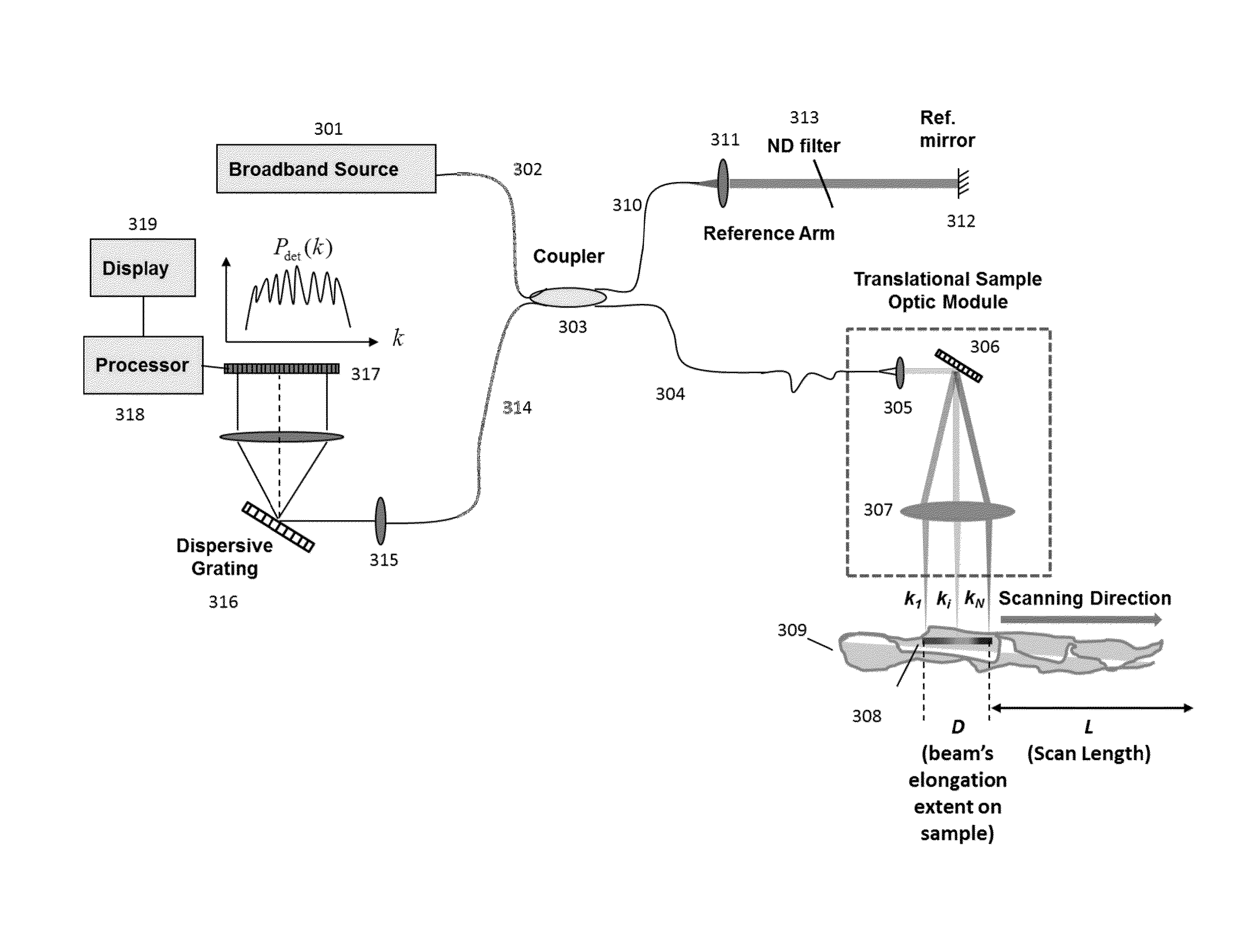
Figure 1 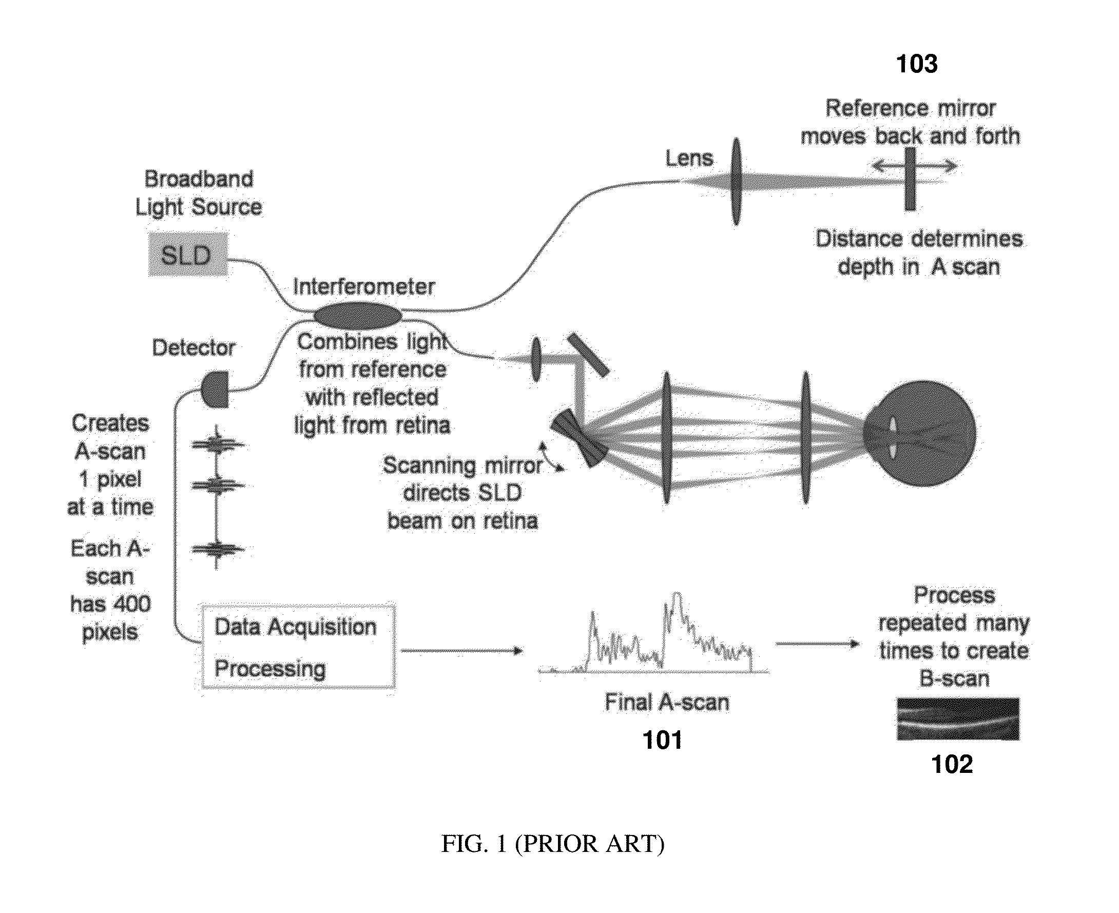
Figure 2 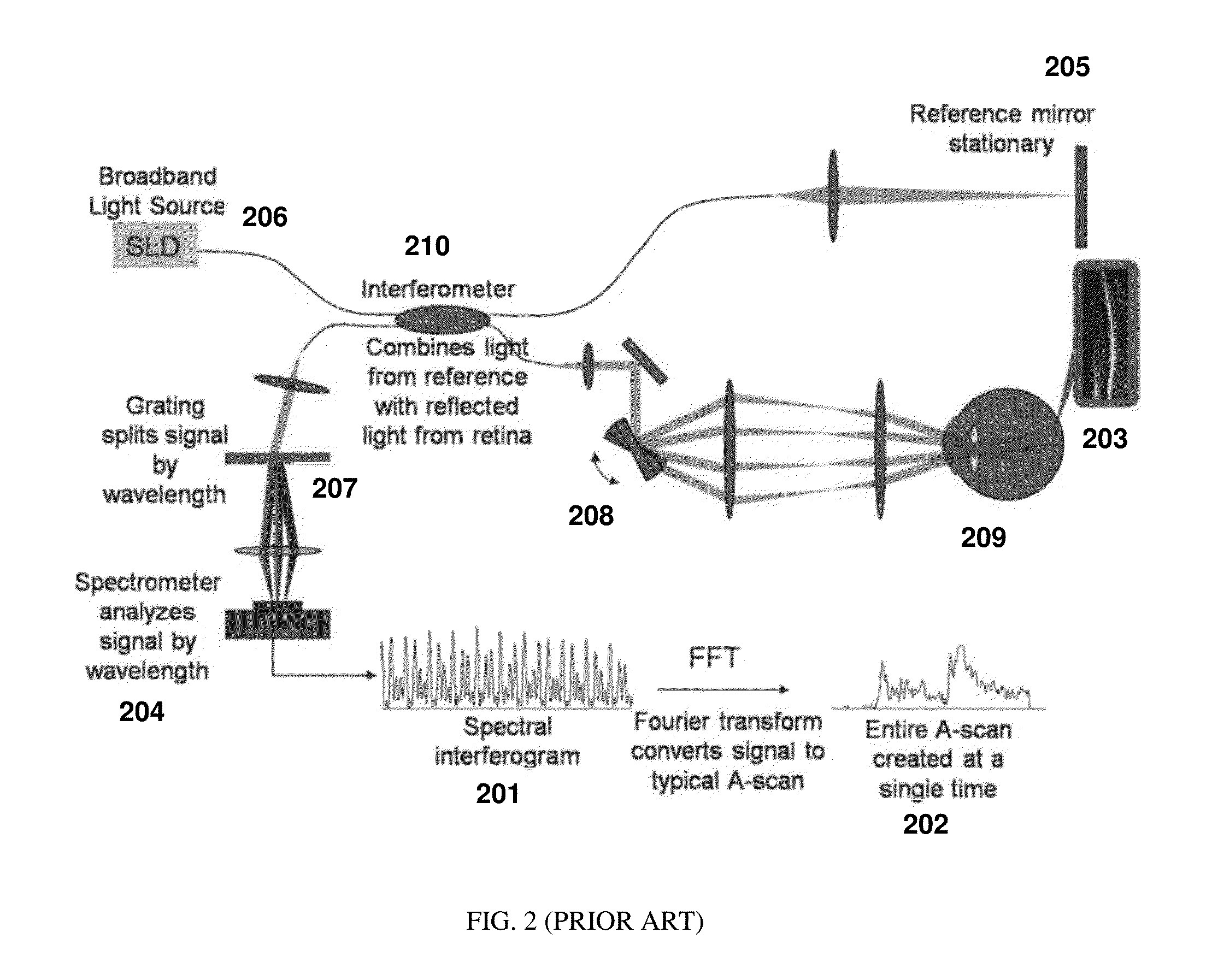
Figure 3
Abstract
Description
PRIORITY
[0001] The present application claims priority to U.S. Provisional Application Ser. No. 62 / 020,556 filed Jul. 3, 2014 the contents of which are hereby incorporated by reference.TECHNICAL FIELD
[0002] The present application relates to the field of optical coherence tomography. In particular, the present application describes embodiments related to producing and processing partial spectrum A-scans with optical coherence tomographic instrumentation.BACKGROUND
[0003] Optical Coherence Tomography (OCT) is an interferometric technique that can provide images of samples including tissue structure on the micrometer scale in situ and in real time (Huang, D. et al., Science 254, 1178-81, 1991). OCT is based on the principle of low coherence interferometry (LCI) and determines the scattering profile of a sample along the OCT beam by detecting the interference of light reflected from a sample and a reference beam (Fercher, A. F. et al., Opt. Lett. 13, 186, 1988). Each scattering profile in ...
Examples
Embodiment Construction
[0021]A basic concept of the present application is to spectrally disperse or de-multiplex illumination of the incident light at the sample, move the spectrally dispersed light along the spatial direction of dispersion and spectrally resolve the detection of the light interference. The broadband light from the source is distributed spectrally on the sample, and each acquisition simultaneously provides partial spectral interference information from multiple locations in the sample. Thus for a given spatial point, a single observation will be of a partial spectrum A-scan. When multiple partial spectrum A-scan observations are made at the same point by shifting the spectrum of light on to the tissue, the point will be illuminated by the entire broadband spectrum of the light source over time, thereby making it possible to construct a full axial resolution A-scan.
[0022]Spectral spreading of the incident beam on a sample makes it possible to increase incident power within safe limits. He...