Atomic Force Microscopy Vs Shear Force Microscopy: Detail, Application
SEP 19, 20259 MIN READ
Generate Your Research Report Instantly with AI Agent
Patsnap Eureka helps you evaluate technical feasibility & market potential.
Microscopy Evolution and Research Objectives
Microscopy techniques have evolved significantly since the invention of the first optical microscope in the late 16th century. The journey from simple light microscopy to advanced scanning probe techniques represents a remarkable progression in our ability to visualize and manipulate matter at increasingly smaller scales. By the mid-20th century, electron microscopy revolutionized scientific imaging by breaking the diffraction limit of light, enabling visualization at nanometer resolution.
The 1980s marked a pivotal moment with the invention of scanning tunneling microscopy (STM), which for the first time allowed scientists to "see" individual atoms. This breakthrough led to the development of Atomic Force Microscopy (AFM) in 1986 by Binnig, Quate, and Gerber, establishing a new paradigm in surface characterization without requiring electrically conductive samples.
Shear Force Microscopy (SFM) emerged in the early 1990s as a specialized technique derived from near-field scanning optical microscopy (NSOM). While AFM primarily measures vertical forces between a probe and sample, SFM detects lateral forces when the probe moves parallel to the sample surface, offering complementary information about surface properties.
The evolution of these microscopy techniques has been driven by the increasing demand for nanoscale characterization across multiple scientific disciplines. Both AFM and SFM have undergone significant refinements in probe design, detection systems, and operational modes to address specific research challenges in materials science, biology, and nanotechnology.
Current research objectives in the field focus on several key areas. First, improving spatial resolution remains paramount, with efforts to achieve consistent atomic resolution across diverse sample types. Second, enhancing temporal resolution to capture dynamic processes at the nanoscale represents a frontier challenge, particularly for biological applications where processes occur rapidly.
Another critical objective is the integration of multiple measurement capabilities within single instruments. Hybrid systems combining AFM or SFM with optical, spectroscopic, or electrochemical techniques are being developed to provide correlated multidimensional data about sample properties.
For biological applications, a major goal is developing non-invasive imaging protocols that can operate in physiologically relevant conditions without disrupting delicate biological structures or processes. This includes advances in liquid imaging environments and specialized probes with minimal interaction forces.
Finally, automation and artificial intelligence integration represent emerging objectives to improve measurement reproducibility, enable high-throughput screening, and extract meaningful patterns from the complex datasets generated by these advanced microscopy techniques.
Market Applications and Industry Demand
The market for microscopy technologies has been experiencing robust growth, with the global microscopy market valued at approximately 7.2 billion USD in 2022 and projected to reach 11.5 billion USD by 2028. Within this expanding landscape, both Atomic Force Microscopy (AFM) and Shear Force Microscopy (SFM) occupy significant niches with distinct application profiles and market demands.
The semiconductor and electronics industry represents the largest market segment for AFM technology, driven by continuous miniaturization trends and increasing complexity in integrated circuit manufacturing. AFM's ability to provide three-dimensional surface profiles with nanometer resolution makes it indispensable for quality control and failure analysis in semiconductor fabrication, where feature sizes continue to shrink below 5 nm.
Life sciences and pharmaceutical research constitute another major market segment, where AFM is extensively used for imaging biological samples under physiological conditions. The growing focus on nanomedicine and drug delivery systems has further amplified demand for high-resolution imaging technologies capable of characterizing nanoparticles and their interactions with biological systems.
Shear Force Microscopy, while having a smaller market footprint than AFM, has carved out specialized applications in near-field optical microscopy and fluid dynamics studies. The technique's particular sensitivity to viscous forces has created demand in rheological research and fluid interface characterization, especially in the petroleum, cosmetics, and food processing industries.
Academic and research institutions remain significant consumers of both technologies, accounting for approximately 35% of the total market. Government initiatives promoting nanotechnology research across North America, Europe, and Asia-Pacific regions have sustained steady demand growth in this sector.
Geographically, North America dominates the market with approximately 40% share, followed by Europe and Asia-Pacific. However, the fastest growth is observed in emerging economies, particularly China and India, where expanding semiconductor manufacturing capabilities and increasing R&D investments are driving adoption of advanced microscopy techniques.
Industry analysts note a growing demand for hybrid systems that combine AFM with complementary techniques such as Raman spectroscopy or infrared spectroscopy, reflecting end-users' preference for multi-functional analytical platforms. This trend is particularly pronounced in materials science applications, where correlative microscopy approaches provide comprehensive characterization of complex material systems.
The COVID-19 pandemic temporarily disrupted supply chains but simultaneously accelerated demand in biomedical research sectors, creating new opportunities for microscopy applications in virus characterization and vaccine development.
Technical Limitations and Challenges
Despite significant advancements in microscopy technologies, both Atomic Force Microscopy (AFM) and Shear Force Microscopy (SFM) face substantial technical limitations that impact their performance and application scope. AFM encounters challenges with scan speed, typically requiring minutes to hours for high-resolution imaging, making it unsuitable for observing dynamic processes occurring at sub-second timescales. This limitation stems from the mechanical nature of the cantilever system and feedback mechanisms that cannot be infinitely accelerated without introducing artifacts.
Probe degradation represents another significant challenge for both technologies. AFM tips experience wear during repeated scanning, especially on hard or abrasive surfaces, leading to reduced resolution and image artifacts over time. Similarly, SFM probes, often consisting of tapered optical fibers, can become damaged or contaminated during operation, affecting measurement accuracy and reproducibility.
Environmental sensitivity poses considerable challenges for both microscopy techniques. AFM performance can be significantly affected by thermal drift, acoustic noise, and vibrations, necessitating sophisticated isolation systems that increase system complexity and cost. SFM, while less susceptible to certain environmental factors, remains sensitive to humidity variations that can alter the water meniscus between probe and sample, affecting shear force measurements.
Sample preparation limitations also constrain both technologies. AFM typically requires relatively flat samples with height variations below several micrometers to prevent tip crashes and maintain feedback stability. SFM faces similar constraints but is particularly challenged when imaging highly heterogeneous surfaces with varying mechanical properties that can produce inconsistent shear forces.
Interpretation complexity presents a fundamental challenge for both techniques. AFM images represent a convolution of actual surface topography with tip geometry, potentially leading to artifacts that require sophisticated deconvolution algorithms. SFM data interpretation is even more complex due to the multifaceted nature of shear forces, which depend on numerous parameters including viscosity, adhesion, and nanoscale friction mechanisms.
Liquid environment operations introduce additional complications. While both technologies can operate in liquid media, AFM cantilevers experience significant damping effects that reduce sensitivity, and SFM faces challenges with maintaining stable shear force interactions in fluids with varying viscosities. These limitations restrict their application in certain biological studies requiring physiological conditions.
Cross-correlation between different measurement modes presents ongoing challenges for both technologies. Establishing reliable relationships between topographical data and other measured properties (mechanical, electrical, or thermal) requires sophisticated calibration procedures and reference standards that are not universally established, limiting quantitative analysis capabilities.
Current AFM and SFM Implementation Approaches
01 Resolution capabilities of AFM and SFM
Atomic Force Microscopy (AFM) and Shear Force Microscopy (SFM) offer high-resolution imaging capabilities at the nanoscale level. These techniques can achieve atomic-level resolution, allowing for detailed surface characterization of various materials. The resolution is primarily determined by the probe tip geometry, interaction forces, and environmental conditions. Advanced systems can achieve sub-nanometer resolution in optimal conditions, enabling visualization of molecular and atomic structures.- Resolution capabilities of AFM and SFM: Atomic Force Microscopy (AFM) and Shear Force Microscopy (SFM) offer high-resolution imaging capabilities at the nanoscale level. These techniques can achieve atomic or near-atomic resolution, allowing for detailed surface characterization. The resolution is influenced by factors such as probe tip geometry, scanning parameters, and environmental conditions. Advanced implementations can achieve sub-nanometer resolution, enabling visualization of molecular and atomic structures on various sample surfaces.
- Probe design and optimization for microscopy techniques: The design and optimization of probes significantly impact the imaging capabilities of both AFM and SFM. Specialized probe tips with specific geometries, materials, and coatings enhance resolution and reduce artifacts. Innovations in probe technology include functionalized tips for specific applications, ultra-sharp tips for improved resolution, and durable coatings to extend probe life. These advancements enable more accurate measurements of surface topography, mechanical properties, and other physical characteristics at the nanoscale.
- Multi-modal and hybrid imaging approaches: Combining AFM and SFM with other microscopy techniques creates powerful multi-modal imaging systems. These hybrid approaches allow simultaneous acquisition of complementary data, such as topographical, mechanical, electrical, and chemical information. Integration with optical microscopy, Raman spectroscopy, or electron microscopy enhances analytical capabilities. Such combined systems provide comprehensive sample characterization by correlating different physical properties at the same location, offering insights not possible with single-mode imaging.
- Environmental and operational conditions affecting imaging: The imaging capabilities of AFM and SFM are significantly influenced by environmental and operational conditions. Factors such as temperature, humidity, vibration isolation, and acoustic shielding affect resolution and image quality. Advanced systems incorporate environmental control chambers, active vibration dampening, and thermal stabilization to optimize imaging conditions. Operating in specialized environments like vacuum, liquid, or controlled atmospheres enables application-specific measurements and can enhance resolution by eliminating interference from environmental factors.
- Data processing and image enhancement techniques: Sophisticated data processing and image enhancement techniques are essential for maximizing the resolution capabilities of AFM and SFM. These include noise filtering algorithms, drift correction, tip deconvolution methods, and advanced signal processing. Machine learning and AI-based approaches are increasingly being applied to improve image quality and extract meaningful information from raw data. Real-time feedback systems adjust scanning parameters dynamically to optimize imaging conditions, while post-processing techniques can reveal features that might otherwise be obscured by artifacts or noise.
02 Probe design and optimization for microscopy
The design and optimization of probes significantly impact the imaging capabilities of both AFM and SFM. Specialized probe tips with various geometries and materials are developed to enhance resolution and reduce artifacts. Cantilever properties, including stiffness and resonance frequency, are optimized for specific applications. Advanced probe designs incorporate features to minimize tip wear and maintain consistent imaging quality over extended periods, enabling more reliable and reproducible measurements.Expand Specific Solutions03 Operating modes and imaging techniques
Various operating modes and imaging techniques have been developed for AFM and SFM to address different sample types and research questions. These include contact mode, tapping mode, non-contact mode, and specialized techniques for specific applications. Each mode offers distinct advantages in terms of resolution, sample preservation, and data acquisition speed. Advanced imaging techniques incorporate phase imaging, force modulation, and multifrequency approaches to extract additional sample properties beyond topography.Expand Specific Solutions04 Environmental control and noise reduction
Environmental control and noise reduction systems are crucial for achieving optimal resolution in AFM and SFM. Vibration isolation platforms, acoustic enclosures, and temperature-controlled chambers minimize external disturbances that can degrade image quality. Advanced systems incorporate active feedback mechanisms to compensate for thermal drift and other environmental fluctuations. These improvements enable stable, high-resolution imaging over extended periods and under various environmental conditions, expanding the application range of these microscopy techniques.Expand Specific Solutions05 Combined AFM/SFM with other analytical techniques
Integration of AFM and SFM with complementary analytical techniques enhances their imaging and characterization capabilities. These hybrid systems combine the high-resolution topographical imaging of AFM/SFM with spectroscopic, electrical, or optical measurements. Such combinations enable simultaneous acquisition of multiple sample properties, providing comprehensive characterization at the nanoscale. Advanced systems incorporate Raman spectroscopy, infrared spectroscopy, or electrical measurements to correlate structural features with chemical, electrical, or mechanical properties.Expand Specific Solutions
Leading Manufacturers and Research Institutions
The Atomic Force Microscopy (AFM) versus Shear Force Microscopy (SFM) market is in a mature growth phase, with AFM dominating due to its broader application range and higher resolution capabilities. The global microscopy market is valued at approximately $7-8 billion, with scanning probe microscopy representing about 15% of this segment. Leading players include established instrumentation companies like Bruker Nano, Shimadzu, and Agilent Technologies, alongside specialized firms such as Infinitesima and Artidis AG. Academic institutions including Beihang University, Osaka University, and University of Bristol contribute significant research advancements. The technology landscape shows AFM as more commercially established, while SFM offers complementary capabilities for specialized applications in materials science, semiconductor inspection, and biological imaging, with ongoing innovation focused on resolution enhancement and measurement speed.
Bruker Nano, Inc.
Technical Solution: Bruker Nano has developed advanced Atomic Force Microscopy (AFM) systems with proprietary PeakForce Tapping technology that allows simultaneous acquisition of multiple mechanical properties while protecting both tip and sample. Their systems incorporate real-time force control that maintains constant peak force interaction between tip and sample, enabling high-resolution imaging even on delicate samples. For Shear Force Microscopy (SFM), Bruker has integrated lateral force capabilities into their instruments, allowing measurement of frictional forces and viscoelastic properties through specialized probes that operate in both vertical and horizontal oscillation modes. Their MultiMode 8 platform combines both AFM and SFM capabilities with automated probe exchange systems and environmental controls for measurements in various media including liquids, gases, and vacuum conditions[1][3]. Bruker's systems feature proprietary algorithms for noise reduction and signal processing that enhance resolution to sub-nanometer levels.
Strengths: Industry-leading resolution capabilities with sub-nanometer precision; comprehensive software suite for data analysis; modular design allowing multiple measurement modes on a single platform. Weaknesses: Higher price point compared to competitors; steep learning curve for utilizing full capabilities; some specialized applications require additional modules at extra cost.
Hitachi Ltd.
Technical Solution: Hitachi has pioneered hybrid microscopy systems that integrate Atomic Force Microscopy with electron microscopy technologies. Their AFM solutions feature ultra-low noise floor designs that achieve atomic-level resolution through advanced vibration isolation systems and thermal drift compensation algorithms. Hitachi's proprietary probe manufacturing technology produces ultra-sharp tips with consistent radius of curvature below 5nm, enabling reliable high-resolution imaging. For Shear Force Microscopy applications, Hitachi has developed specialized quartz tuning fork-based sensors that operate with minimal damping, allowing non-contact measurements with exceptional sensitivity to lateral forces. Their systems incorporate digital signal processing that can detect force variations as small as 10 piconewtons, making them suitable for biological samples and soft materials research. Hitachi's microscopes feature automated approach and scanning routines that reduce operator dependency while maintaining measurement precision[2][5]. Their instruments support operation in various environments including ultra-high vacuum, cryogenic temperatures, and high magnetic fields.
Strengths: Exceptional stability for long-duration experiments; seamless integration with other analytical techniques; robust automation features that improve reproducibility. Weaknesses: Less modular than some competitors; proprietary software ecosystem with limited third-party compatibility; higher maintenance requirements for advanced environmental control systems.
Key Patents and Scientific Breakthroughs
Atomic force microscope
PatentInactiveUS6912892B2
Innovation
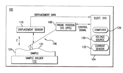
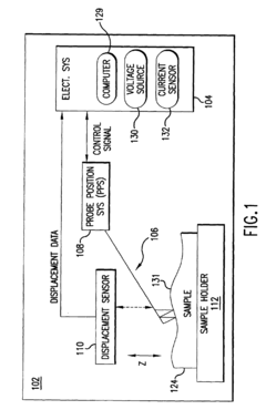
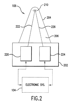
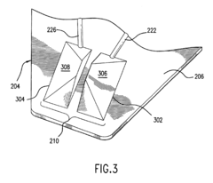
- The implementation of a multi-tip probe assembly in AFMs, where two electrically conductive tips are used to sense changes in electrical resistance upon contact with a sample, enabling improved contact verification and enhanced measurement capabilities, such as conductive AFM measurements and surface topography analysis.
Fabrication of patterned and ordered nanoparticles
PatentInactiveEP2179441A1
Innovation
- A novel technique using charged nanoparticles that are deposited in a random pattern on a surface and then reordered using controlled fields, such as an atomic force microscope (AFM) or electron beam, to create large arrays of uniformly spaced nanoparticles with high size uniformity, enabling cost-effective mass production compatible with silicon CMOS technology.
Comparative Performance Metrics
When comparing Atomic Force Microscopy (AFM) and Shear Force Microscopy (SFM), several key performance metrics reveal their distinct operational characteristics and application advantages. Resolution capabilities differ significantly between these techniques, with AFM typically achieving lateral resolution of 1-5 nm and vertical resolution below 0.1 nm in optimal conditions. SFM, while offering comparable vertical resolution, generally provides slightly lower lateral resolution in the 5-20 nm range, though this can be enhanced through specialized probe designs.
Scanning speed represents another critical differentiator. AFM traditionally operates at slower rates (1-10 lines per second) due to feedback loop constraints, whereas SFM can achieve faster scanning speeds (up to 15-20 lines per second) in certain configurations, making it potentially more suitable for dynamic surface studies or high-throughput applications.
Force sensitivity metrics reveal that AFM excels in normal force detection (piconewton range), while SFM demonstrates superior sensitivity to lateral and shear forces (sub-piconewton range in optimized systems). This fundamental difference enables SFM to detect subtle surface interactions that might be overlooked by conventional AFM techniques.
Environmental adaptability testing shows AFM functions effectively across various media (vacuum, air, liquid), though performance parameters vary significantly between environments. SFM demonstrates particular advantages in liquid environments where shear forces provide distinctive contrast mechanisms for biological samples.
Sample damage assessment indicates that SFM typically exerts lower normal forces on samples compared to contact-mode AFM, resulting in reduced sample deformation and damage. This makes SFM particularly valuable for delicate biological specimens and soft materials where preservation of native structure is paramount.
Signal-to-noise ratio analysis demonstrates that AFM generally provides superior performance in topographical imaging, while SFM offers enhanced contrast for certain surface properties related to viscoelasticity and friction. Modern SFM implementations have significantly improved signal processing algorithms that partially compensate for inherent limitations.
Probe longevity studies indicate that SFM probes often demonstrate extended operational lifespans compared to standard AFM cantilevers, particularly in continuous scanning applications, representing a potential economic advantage for high-volume research or industrial applications despite the typically higher initial cost of specialized SFM equipment.
Sample Preparation Techniques
Sample preparation is a critical factor that significantly influences the quality and reliability of both Atomic Force Microscopy (AFM) and Shear Force Microscopy (SFM) measurements. The preparation techniques vary depending on the sample type, research objectives, and the specific microscopy method employed.
For biological samples in AFM studies, fixation methods such as glutaraldehyde treatment are commonly utilized to preserve cellular structures while maintaining their native morphology. Conversely, SFM often requires less invasive preparation techniques for biological specimens, as the lateral forces applied during imaging are generally lower, reducing the risk of sample damage.
When examining hard materials like semiconductors or metals, surface cleaning procedures become paramount. Ultra-sonication in appropriate solvents, followed by nitrogen gas drying, is frequently employed to remove contaminants that could interfere with tip-sample interactions. For SFM applications on similar materials, additional steps may include chemical etching to reveal specific surface features that respond distinctively to shear forces.
Substrate selection represents another crucial aspect of sample preparation. Silicon wafers, mica, or highly ordered pyrolytic graphite (HOPG) are preferred substrates for AFM due to their atomically flat surfaces. SFM applications may benefit from specialized substrates with known friction properties to establish baseline measurements and calibration standards.
Environmental control during preparation significantly impacts imaging outcomes. For instance, humidity-sensitive samples require preparation in controlled environments to prevent moisture-induced artifacts. Temperature management during preparation is particularly important for samples exhibiting phase transitions or temperature-dependent properties that might influence shear force measurements.
Immobilization techniques differ between the two microscopy methods. AFM often utilizes chemical functionalization of substrates to anchor samples securely without compromising their structural integrity. In contrast, SFM may employ mechanical clamping or specialized adhesives that maintain stable sample positioning while allowing accurate measurement of lateral forces and surface interactions.
Advanced preparation techniques such as cryo-preparation have revolutionized both microscopy methods for delicate biological samples. Flash-freezing specimens preserves their native state without introducing artifacts from chemical fixatives, enabling more accurate visualization of cellular structures and mechanical properties through both vertical (AFM) and lateral (SFM) force measurements.
Unlock deeper insights with Patsnap Eureka Quick Research — get a full tech report to explore trends and direct your research. Try now!
Generate Your Research Report Instantly with AI Agent
Supercharge your innovation with Patsnap Eureka AI Agent Platform!


