Blood vessel extracting method

An extraction method, blood vessel technology, applied in the field of medical tomography image processing, to achieve the effect of good robustness and fast extraction of blood vessels

Active Publication Date: 2014-11-26
SHANGHAI UNITED IMAGING HEALTHCARE
View PDF4 Cites 13 Cited by
  • Summary
  • Abstract
  • Description
  • Claims
  • Application Information

AI Technical Summary

Problems solved by technology

[0007] The problem solved by the present invention is to provide a blood vessel extraction method to solve the problem of head and neck blood vessel extraction and segmentation

Method used


Image

  • Blood vessel extracting method
  • Blood vessel extracting method
  • Blood vessel extracting method

Examples


Embodiment Construction

[0024] In the following description, numerous specific details are set forth in order to provide a thorough understanding of the present invention. However, the present invention can be implemented in many other ways different from those described here, and those skilled in the art can make similar extensions without violating the connotation of the present invention, so the present invention is not limited by the specific implementations disclosed below.

[0025] Secondly, the present invention is described in detail by means of schematic diagrams. When describing the embodiments of the present invention in detail, for convenience of explanation, the schematic diagrams are only examples, which should not limit the protection scope of the present invention.

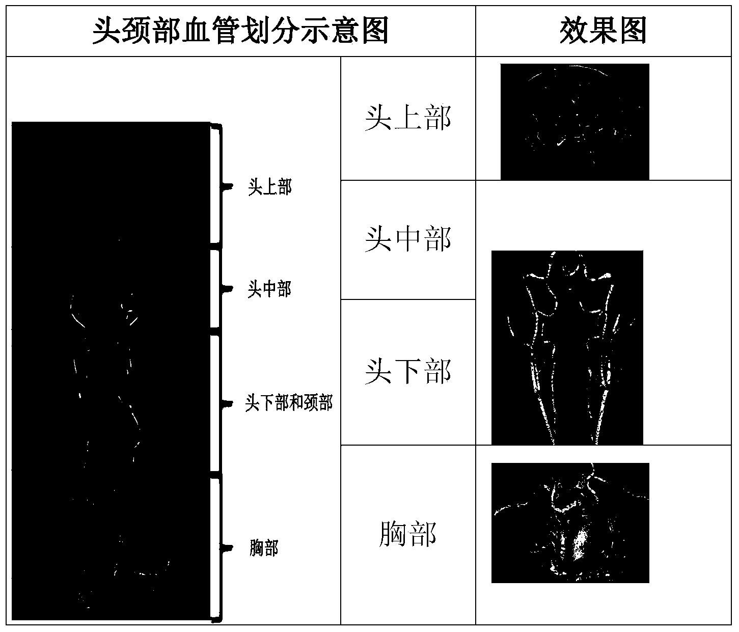
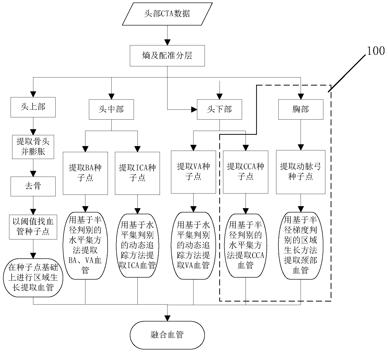
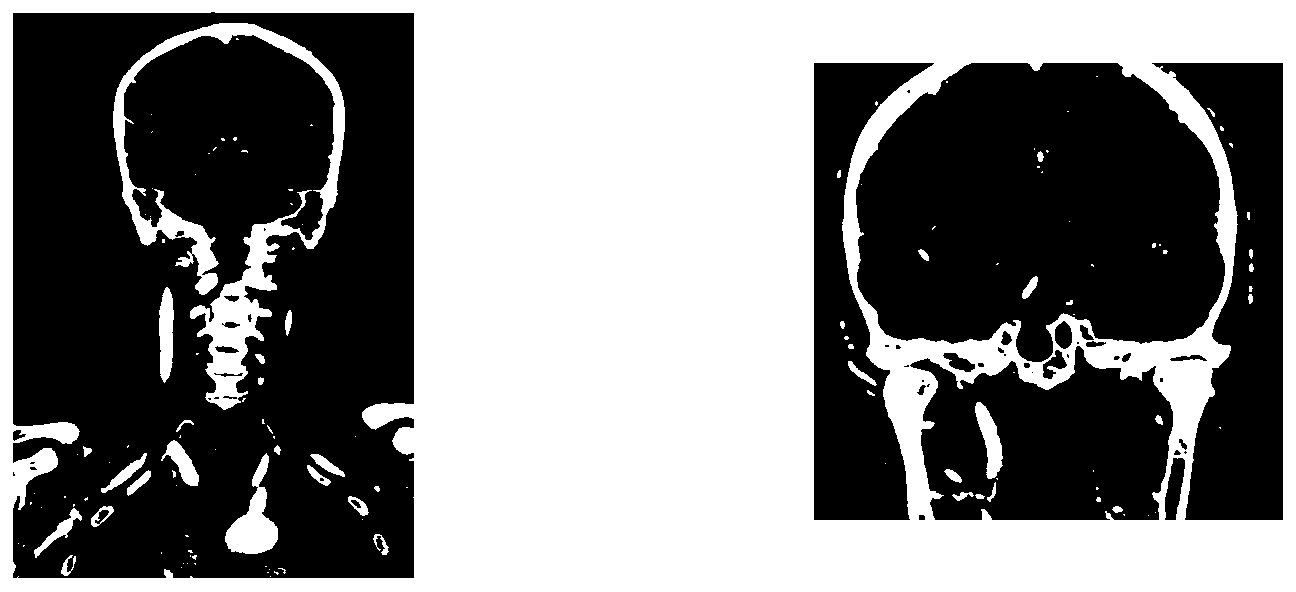
[0026] In order to solve the problem of head and neck blood vessel extraction and segmentation, the present invention provides a blood vessel extraction method, which includes providing head and neck CT contrast data, and ...


PUM


Abstract

The invention provides a blood vessel extracting method comprising the steps of providing head and neck CT angiography data, dividing the head and neck CT angiography data into a plurality of parts, and adopting corresponding vessel extraction algorithms to perform blood vessel extraction on the plurality of parts. Considering that vessels of different parts of the head and neck region differ greatly in morphology, the head and neck region is divided into four blocks in the method, and extraction of the blood vessels of the head and neck region is realized by combining a region growing algorithm, a level set algorithm and a dynamic tracking algorithm. The method has good robustness, is high in blood vessel extraction speed, and is applicable to data of different manufacturers.

Description

technical field [0001] The invention relates to a processing method of medical tomography images, in particular to a blood vessel extraction method. Background technique [0002] Computed Tomography Ang iography (CTA for short) is one of the important methods for diagnosing vascular diseases at present. However, since the blood vessels in the CTA image overlap with the CT values ​​of other tissues such as bones, extracting the blood vessels from other tissues is a key step in angiography. [0003] American literature 1: P.T.Vieco, W.P.Shuman, G.F.Alsofrom, C.E.Gross.Detection of circle of Willis aneurysms in patients with acute subarachnoid hemorrhage: a comparison of CT angiography and digital subtraction angiography.AJR vol.165no.2425-930)(19 In the blood vessel silhouette method provided, the patient needs to scan the same part twice (scanning without contrast agent injection and scanning with contrast agent injection respectively) to obtain non-CTA images and CTA images...

Claims


Application Information

Patent Timeline
Patent Type & Authority Applications(China)
IPC IPC(8): G06T7/00G06T15/00
Inventor 毛玉妃王晓东李程
Owner SHANGHAI UNITED IMAGING HEALTHCARE
Why Eureka
Try Eureka