Medical image Graph Cut segmentation method based on statistical shape model

A technology of statistical shapes and medical images, applied in the field of medical imaging, can solve the problems of inaccurate segmentation, inaccurate segmentation and low efficiency.

Active Publication Date: 2017-03-08
NORTHWEST UNIV
View PDF2 Cites 26 Cited by
  • Summary
  • Abstract
  • Description
  • Claims
  • Application Information

AI Technical Summary

Problems solved by technology

[0006] The purpose of the present invention is to propose a segmentation method based on a statistical shape model and a Graph Cut method for the problems of inaccurate segmentation and low efficiency in the above-mentioned medical image organ segmentation.
Aiming at the problem that the Graph Cut segm

Method used


Image

  • Medical image Graph Cut segmentation method based on statistical shape model
  • Medical image Graph Cut segmentation method based on statistical shape model
  • Medical image Graph Cut segmentation method based on statistical shape model

Examples


Embodiment Construction

[0081] Various details involved in the technical solution of the present invention will be described in detail below in conjunction with the accompanying drawings. It should be pointed out that the described embodiments are only intended to facilitate the understanding of the present invention, rather than limiting it in any way.

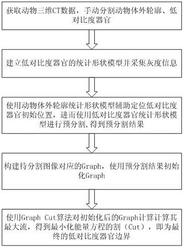
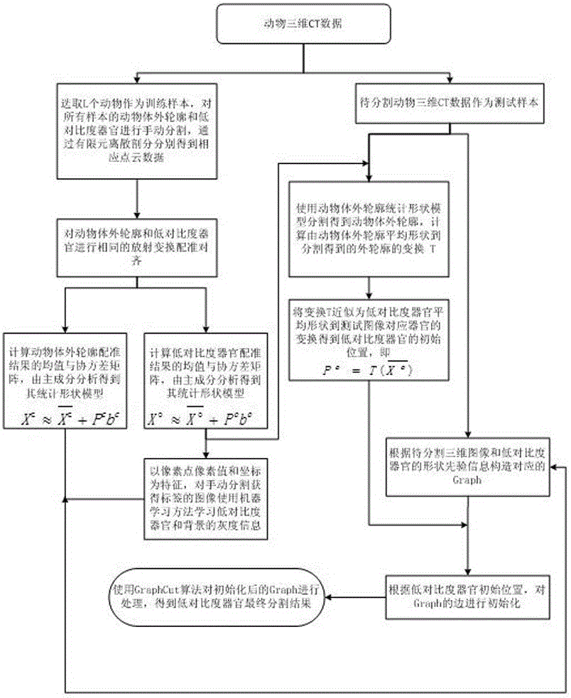
[0082] In this example, mouse kidney was used as the segmentation target, but it is not limited thereto. The framework of the embodiment is attached figure 1 As shown, the detailed process is attached figure 2 shown.

[0083] Step 1: Obtain mouse CT tomographic data

[0084] Fix the experimental mice injected with the contrast agent on the imaging table of the Micro-CT imaging system, adjust the positions of the X-ray tube, the rotating table and the X-ray flat panel detector so that the centers of the three are in a straight line, and perform 360-degree imaging on the mice. High-degree irradiation scanning, acquisition of projection data, and ...


PUM


Abstract

The invention discloses a medical image Graph Cut segmentation method based on a statistical shape model. The medical image Graph Cut segmentation method mainly solves the problem that low-contrast organs cannot be effectively segmented in a medical image in the prior art, and is implemented by the steps of: (1) establishing the statistical shape model of the low-contrast organs, and acquiring grey scale information; (2) pre-segmenting the low-contrast organs; (3) initialization Graph; (4) and segmenting the low-contrast organs. The medical image Graph Cut segmentation method based on the statistical shape model adds priori knowledge of shapes of organs on the basis of rapid segmentation by adopting a Graph Cut algorithm, reduces the possibilities of over-segmentation and under-segmentation, determines initial positions of the low-contrast organs according to relative relationship between animal organs and animal in-vitro contours, improves the segmentation efficiency, and is a rapid and effective organ segmentation method.

Description

technical field [0001] The invention belongs to the field of medical imaging and relates to a statistical contour model and graph cut segmentation. Background technique [0002] As a technology that can provide human body structure information in a non-invasive manner, medical image segmentation is the first step in the analysis and visualization of medical image data. Pathology location, tracking disease progression, and determining radiation therapy dose or surgery size. Therefore, medical image segmentation is a key step in the computer-assisted therapy system, and the accuracy of segmentation directly affects the effect of auxiliary therapy. [0003] However, accurate segmentation of medical images faces many challenges. First, many anatomical structures are non-uniform under the spatial overlap of pixel or voxel gray levels. Secondly, there are low-contrast parts in medical images. For example, the edges of kidneys and hearts are difficult to distinguish in CT images...

Claims


Application Information

Patent Timeline
IPC IPC(8): G06T7/00G06T7/11G06T7/38
Inventor 赵凤军陈雁蓉贺小伟贺小慧高培何雪磊孙飞飞曹欣易黄建侯榆青
Owner NORTHWEST UNIV
Why Eureka
Try Eureka