Uterine oviduct angiography pipe

A technology for fallopian tubes and implanting uterus, which is applied in the direction of catheters, drug devices, and other medical devices. It can solve problems such as cumbersome operation procedures, and achieve the effects of convenient use, low cost, and easy promotion

Pending Publication Date: 2017-08-04
NANTONG YIKAI MEDICAL DEVICES CO LTD
View PDF5 Cites 1 Cited by
  • Summary
  • Abstract
  • Description
  • Claims
  • Application Information

AI Technical Summary

Problems solved by technology

Similarly, because of the need to enter the uterine cavity, although the examination is performed in the radiology department, this

Method used


Image

  • Uterine oviduct angiography pipe

Examples


Embodiment Construction

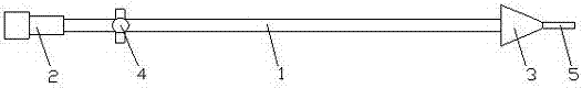
[0015] Such as figure 1 A hysterosalpingography tube as shown comprises a catheter 1, a connector 2 and an endocervical endocervical ostium blocking end 3, one end of the catheter 1 is fixedly connected to the adapter 2, and the other end of the catheter is fixed to the endocervical endocervical ostium blocking end 3 Connecting, a valve 4 is fixedly arranged on the catheter 1 close to the joint 2, the end of the endocervical canal blocking the outer opening 3 is a conical body structure, and the top of the end of the endocervical opening blocking end 3 is fixedly provided with a front-end catheter 5, and the inside of the front-end catheter 5 The cavity communicates with the cavity in the endocervix endocervix 3 and the cavity in the catheter 1.

[0016] Insert the front-end catheter into the external opening of the cervical canal, and the external opening of the cervical canal is blocked by the end 3 to block the cervical canal. After adjusting the proper position, it is fixe...


PUM


Abstract

The invention discloses a uterine oviduct angiography pipe. The pipe comprises a conduit, a connector and a cervical canal external opening obstructing end, one end of the conduit is fixedly connected with the connector, the other end of the conduit is fixedly connected with the cervical canal external opening obstructing end, the portion, close to the connector, of the conduit is further fixedly provided with a valve, the cervical canal external opening obstructing end is of a conical structure, the top of the cervical canal external opening obstructing end is fixedly provided with a front end conduit, and a cavity of the front end conduit is communicated with a cavity of the cervical canal external opening obstructing end and a cavity of the conduit. The uterine oviduct angiography pipe has the advantages of being simple in structure, convenient to use, low in cost and easy to popularize; the adopted cervical canal external opening obstructing end and the front end conduit are easier to fix at an external cervical orifice and do not need to enter a uterine cavity, cervical clamps are not needed to pull cervix uterus, the operation can be completed merely by one worker, the uterine oviduct angiography pipe is safe and reliable in use, is not prone to loosening, and thus liquid leakage can be prevented; the adopted valve can improve the convenience in use, and improve the working efficiency.

Description

[0001] count field [0002] The invention relates to the technical field of medical equipment, in particular to a hysterosalpingography tube. Background technique [0003] At present, due to the influence of environment, social factors and lifestyle, the incidence of tubal infertility is increasing year by year. Hysterosalpingography (HSG) and fluid flow often use double-lumen balloon catheters or metal rod-shaped contrast instruments, and all instruments need to enter the uterine cavity, which increases the patient's pain and chances of infection. In addition, artificial filling defects in the uterine cavity caused by the filled balloon often cover up uterine cavity lesions and even make one side of the fallopian tube invisible, which significantly affects the diagnosis of uterine cavity and fallopian tube lesions by HSG. [0004] Because the instrument needs to enter the uterine cavity, it is an invasive operation, and the operation time is long, which increases the pain of...

Claims


Application Information

Patent Timeline
IPC IPC(8): A61M31/00A61M25/00
CPCA61M25/0043A61M31/00A61M2210/1433
Inventor 韩志刚田复波秦晓鹏魏先锋
Owner NANTONG YIKAI MEDICAL DEVICES CO LTD
Why Eureka
Try Eureka