High-resolution X-ray imaging device of tissues in human body

A high-resolution, optical imaging technology, applied in the field of medical imaging, can solve problems such as misjudgment and unclear imaging, and achieve the effect of reducing pain, clear imaging, and convenient judgment.

Pending Publication Date: 2018-06-19
陕西鑫威泰生物科技有限公司
View PDF1 Cites 4 Cited by
  • Summary
  • Abstract
  • Description
  • Claims
  • Application Information

AI Technical Summary

Problems solved by technology

Although this patent can extend into the human body and perform radiation imaging on the target area from the body, the imaging is not clear and it is easy to cause misjudgment

Method used


Image

  • High-resolution X-ray imaging device of tissues in human body
  • High-resolution X-ray imaging device of tissues in human body
  • High-resolution X-ray imaging device of tissues in human body

Examples


Embodiment 1

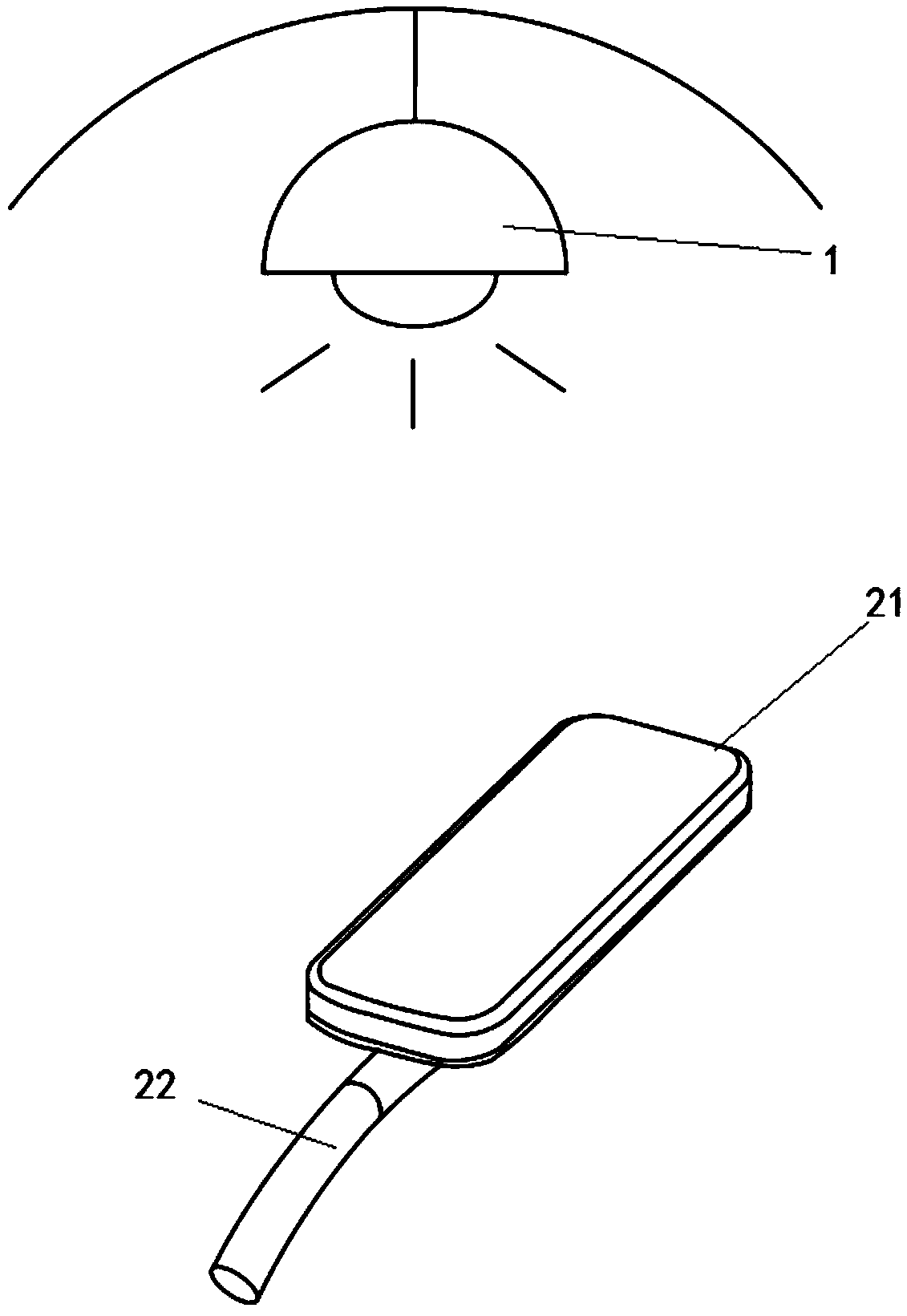
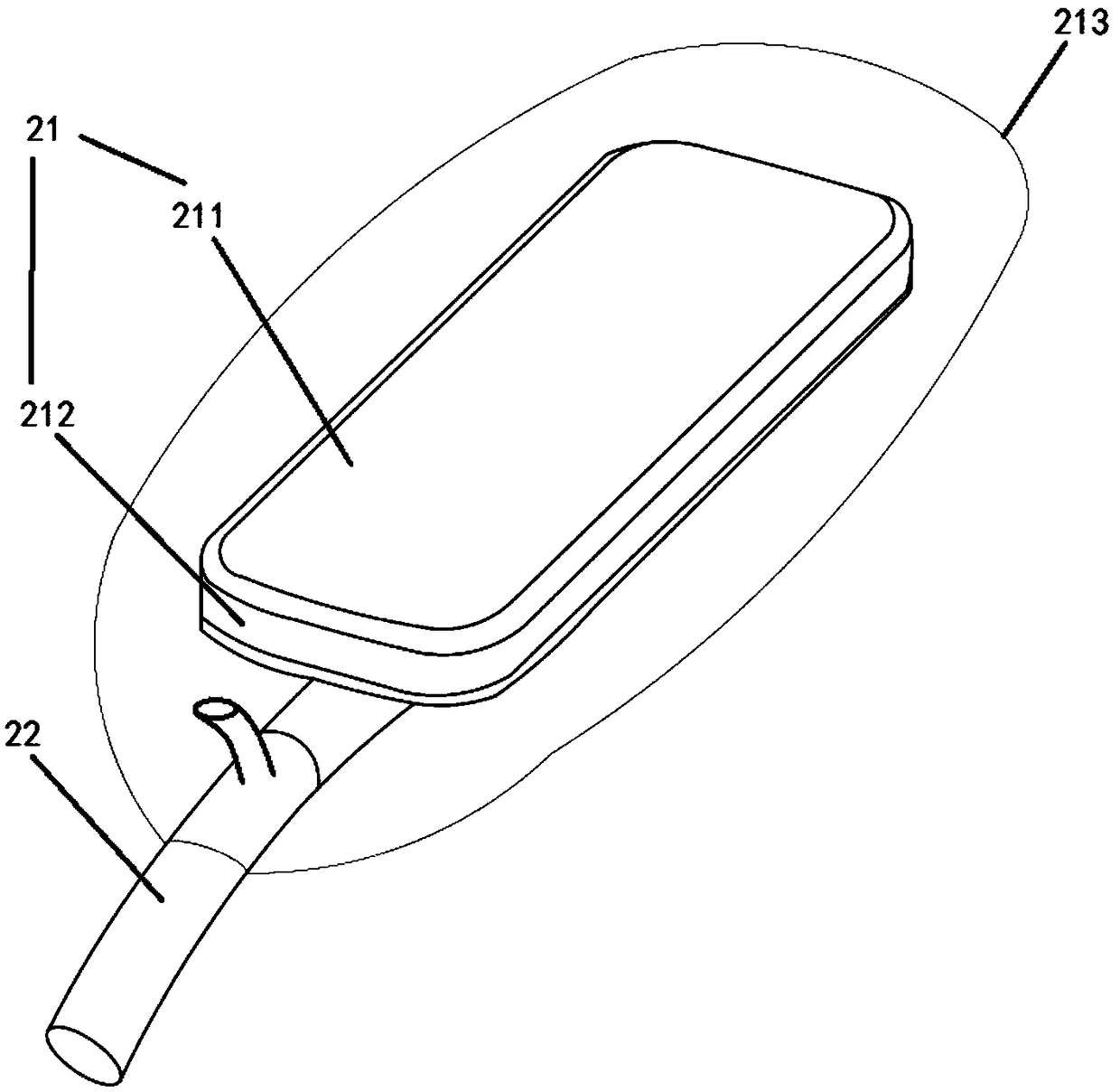
[0032] like figure 2 As shown, in this embodiment, the imaging device 2 is composed of an imaging assembly 21; the imaging assembly 21 is composed of a detection surface a211 and a radiation detector a212, and the bottom surface of the detection surface a211 is fixedly connected to the upper surface of the radiation detector a212 , the radiation detector a212 is coupled with the delivery structure 22; the imaging component 21 is sheathed with a fixed inflatable bag 213 on the outer surface, and the inflatable bag 213 communicates with the inflatable device outside the human body through a hose penetrated by the delivery structure 22.

[0033] The imaging device 2 is set to a size and shape that allows it to be inserted into the patient's body. When in use, it is inserted into the patient's body. The detection surface a211 faces the detection target area, and then the inflatable device is inflated to fix the imaging device 2. The radiation source 1 is turned on, and the radiati...

Embodiment 2

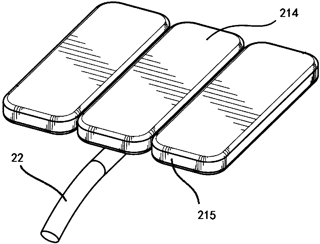
[0035] In this embodiment, the imaging device 2 is composed of three imaging components 21; the imaging component 21 is composed of a detection surface b214 and a radiation detector b215; the adjacent detection surfaces b214 are flexibly connected; the radiation detection The device b 215 is coupled to the delivery structure 22.

[0036] The imaging device 2 is set to a size and shape that allows it to be inserted into the patient's body. After being inserted into the patient's body, the imaging assembly 21 is expanded to increase the area of ​​the detection surface b214, and greatly reduce the truncation artifact caused by the target area being larger than the surface of the imaging device 2. film.

[0037] An electric expansion device can be added between adjacent detection surfaces b214 to achieve a better technical effect.

Embodiment 3

[0039] like image 3 As shown, as a preferred embodiment of Embodiment 2, in this embodiment, the surface of the detection surface b214 is an inflatable air bag, which communicates with the inflatable device outside the human body through the flexible tube penetrated by the delivery structure 22, and adjacent detection The surface b214 is connected.

[0040] When in use, the inflatable device is turned on, and the three detection surfaces b214 are inflated and unfolded. After the use is completed, the gas inside is extracted, and the three detection surfaces b214 are pressed and folded.


PUM


Abstract

The invention discloses a high-resolution X-ray imaging device of tissues in human body. The X-ray imaging device comprises a radiation source (1) for emitting electromagnetic radiation and an imagingdevice (2), wherein the imaging device (2) comprises at least one imaging component (21) and a delivery structure (22) coupled with each imaging component (21); the imaging components (21) are connected with each other, extend into the human body in use, are fixed in a diagnostic target area and cooperates with the radiation source (1) arranged outside the human body to image; the imaging components (21) are coupled with the delivery structure (22). The high-resolution X-ray imaging device of tissues in human body is capable of accurately imaging the tissues in the human body; the auxiliary manner used when the device in the prior art is incapable of imaging is avoided; the pain of the patient is relieved; meanwhile, the imaged images are clear and are free of double images, so that the doctor can conveniently and accurately diagnose.

Description

technical field [0001] The invention relates to the field of medical imaging, in particular to high-resolution X-ray imaging equipment for internal tissues. Background technique [0002] Prostate cancer is a common disease among men, with one in seven men in the United States being diagnosed with prostate cancer in their lifetime. Current detection methods for prostate cancer include digital rectal examination (DRE) and prostate-specific antigen (PSA) screening. However, these screening tools tend to have low accuracy rates due to frequent ambiguous results, false positives and even false negatives. In many cases, because screening results are inaccurate, other methods must be used, such as performing a prostate biopsy and analyzing biopsies for cancer pathology. Biopsies are invasive, expensive, and time-consuming, and increase the risk of complications associated with the procedure, such as infection, urinary / sexual dysfunction, and even death. However, because of the i...

Claims


Application Information

Patent Timeline
Patent Type & Authority Applications(China)
IPC IPC(8): A61B6/00
CPCA61B6/425A61B6/4429A61B6/5282
Inventor 王鸿才郝定均郭华贾广何杰顾雪刘坤张翔张真
Owner 陕西鑫威泰生物科技有限公司
Why Eureka
Try Eureka