Automatic urine protein detection equipment

An automatic detection and urine protein technology, which is applied in the field of medical auxiliary equipment, can solve the problems of deviation of detection results, difficulty in manually judging the amount of the amount, objects, physical conditions and other factors, and achieve the effect of improving accuracy

Active Publication Date: 2020-08-14
THE FIRST AFFILIATED HOSPITAL OF ARMY MEDICAL UNIV
View PDF19 Cites 5 Cited by
  • Summary
  • Abstract
  • Description
  • Claims
  • Application Information

AI Technical Summary

Problems solved by technology

[0002] There are often various proteins in the blood, and some of the proteins will be filtered into the urine in the kidney spheroids, but will be absorbed in the renal tubules and returned to the blood. Therefore, when the kidneys are functioning normally, urine The amount of protein in the urine is small, but when the kidney and the urinary catheter are obstructed, the function of the renal tubules to absorb protein will become poor, so that more protein will enter the urine, so clinically, it is usually detected by detecting the protein in the urine. Protein content to judge kidney and urinary catheter diseases
[0003] At present, the detection of urine protein includes three methods: urine protein qualitative test, urine protein quantitative measurement, and urine protein electrophoresis analysis. Among them, the urine protein qualitative test is usually detected with special test paper for urine protein detection, but this can only show the urine protein during detection. The protein content is easily affected by various factors such as the food eaten by the patient at that time, the physical condition, etc., so the test strip can only make a preliminary judgment. Collect the urine volume of the patient for 24 hours, mix the 24-hour urine, and then detect the urine protein content in the mixed urine
[0004] Since the quantitative measurement is performed on 24h mixed urine, in order to improve the accuracy, it is necessary to ensure that the amount of urine collected each time is the same. The amount of urine collected in a certain time is too much or too little, which makes the test results biased

Method used


Image

  • Automatic urine protein detection equipment
  • Automatic urine protein detection equipment
  • Automatic urine protein detection equipment

Examples


Embodiment 1

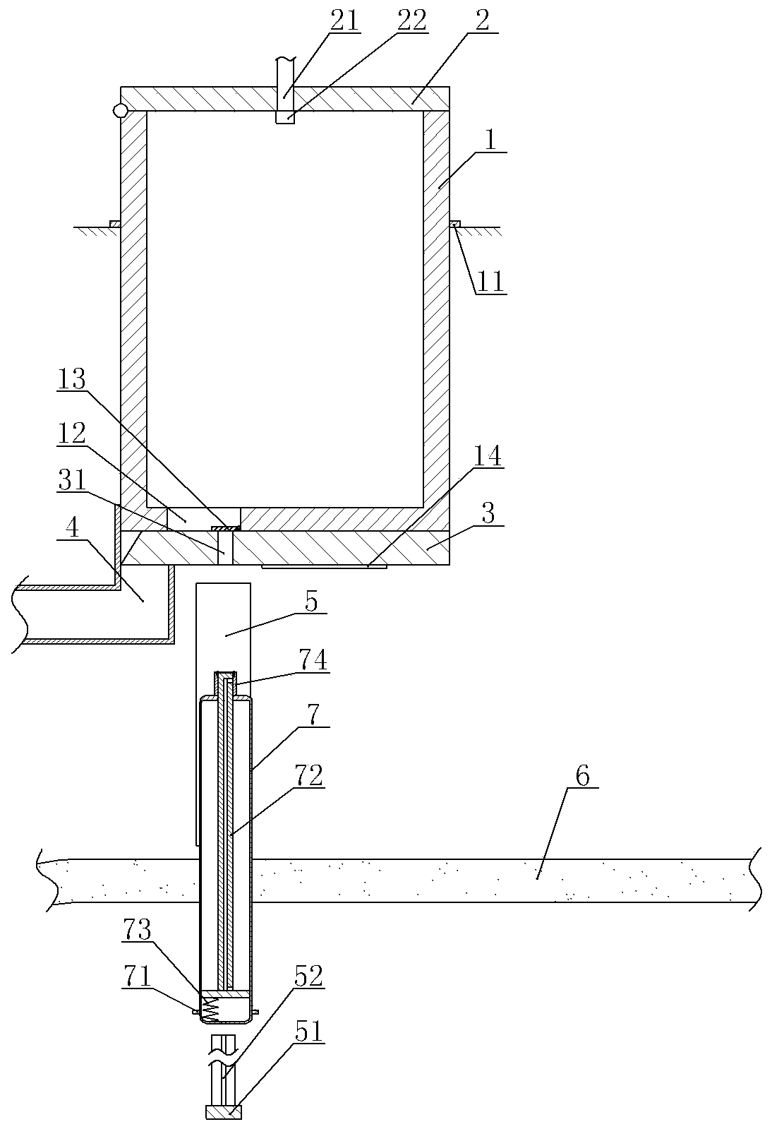
[0039] Urine protein automatic detection equipment, such as figure 1 As shown, it includes a frame, a collection unit and a sample retention unit. The collection unit includes a collection bucket 1 and a sealing cover 3. The outer wall of the collection bucket 1 is welded with a ring-shaped limit block 11. The frame is provided with a mounting hole. The lower end of the collection bucket 1 There is a gap between the side wall of the installation hole to ensure that the lower end of the collection bucket 1 passes through. A cover body 2 is arranged above the collection bucket 1, and the left end of the cover body 2 is hinged on the top of the collection bucket 1 and is covered on the collection bucket 1 to close the opening at the top of the collection bucket 1. A cleaning pipe 21 is provided on the cover 2, and the end of the cleaning pipe 21 runs through the cover 2 and extends into the collection bucket 1. A nozzle 22 is provided under the cover 2, and the nozzle 22 is glued...

Embodiment 2

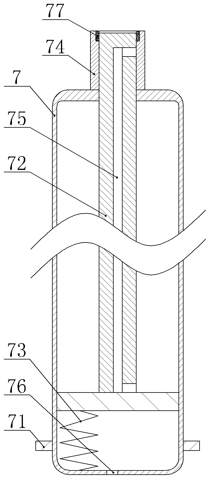
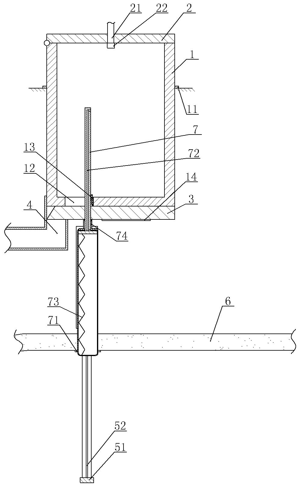
[0053] Such as Figure 4 As shown, the difference from Embodiment 1 is that the sampling channel 31 in this embodiment includes a flared section at the lower end and a necked section at the upper end, the diameter of the flared section is greater than the diameter of the necked section, and the sampling tube 7 slides upwards Finally, the upper end of the detection tube 74 slides into the flared section and abuts against the side wall of the flared section.

[0054] The side wall of the collection bucket 1 is provided with a ring-shaped heating chamber 9, and a heating element 91 is fixed in the heating chamber 9 by bolts. One end away from the heating cavity 9 passes through the cover body 2 and is screwed to the cover body 2 . An air supply unit is fixed on the frame by bolts. Specifically, the air supply unit can be a fan or an air pump. The air supply unit in this embodiment is a fan, and the fan communicates with the heating chamber 9 through a pipe.

[0055] The top of ...


PUM


Abstract

The invention relates to the field of medical auxiliary instruments, and particularly discloses automatic urine protein detection equipment. The equipment comprises a collecting unit and a sample reserving unit. The collecting unit comprises a collecting barrel and a sealing cover, a liquid outlet is formed in the bottom of the collecting barrel, and a sampling channel is formed in the sealing cover. The sample reserving unit comprises a driving part, a rotating part, a pushing part and a plurality of sampling pipes; the plurality of sampling pipes are arranged on the rotating part along the circumferential direction of the rotating part and are detachably connected with the rotating part; a sampling rod is slidably sealed in each sampling pipe; one end of each sampling rod penetrates through one sampling pipe and is in sliding connection with the sampling pipe, the sampling rod and the sampling channel are in sliding seal, and a connecting channel is arranged in each sampling rod; a sampling spring is arranged in each sampling pipe, two ends of each sampling spring are respectively fixed on one sampling pipe and one sampling rod, and the rotating part is positioned below the sampling channels and used for pushing the sampling rods upwards. The equipment can automatically and quantitatively suck urine and store the urine, and the situation that the detection result deviates dueto the fact that the amount of the urine cannot be accurately judged manually is avoided.

Description

technical field [0001] The invention relates to the field of medical auxiliary equipment, in particular to an automatic urine protein detection device. Background technique [0002] There are often various proteins in the blood, and some of the proteins will be filtered into the urine in the kidney spheroids, but will be absorbed in the renal tubules and returned to the blood. Therefore, when the kidneys are functioning normally, urine The amount of protein in the urine is small, but when the kidney and the urinary catheter are obstructed, the function of the renal tubules to absorb protein will become poor, so that more protein will enter the urine, so clinically, it is usually detected by detecting the protein in the urine. Protein content to judge kidney and urinary tract diseases. [0003] At present, the detection of urine protein includes three methods: urine protein qualitative test, urine protein quantitative measurement, and urine protein electrophoresis analysis. ...

Claims


Application Information

Patent Timeline
Patent Type & Authority Applications(China)
IPC IPC(8): G01N1/10G01N33/68B08B9/093B08B13/00
CPCG01N1/10G01N33/6827B08B9/093B08B13/00
Inventor 周悠红白璐郑慧廖素英肖婷
Owner THE FIRST AFFILIATED HOSPITAL OF ARMY MEDICAL UNIV
Why Eureka
Try Eureka