Method and device for the retraction and hemostasis of tissue during crown and bridge procedures

a tissue retraction and hemostasis technology, applied in the field of tissue retraction and hemostasis retraction, can solve the problems of inability to make an impression of the prepared tooth, inconvenient patient treatment, and time-consuming and painful patient treatment, etc., to achieve convenient dentist treatment, improve patient comfort, and improve the effect of operation

Inactive Publication Date: 2005-11-24
CENTRIX
View PDF29 Cites 17 Cited by
  • Summary
  • Abstract
  • Description
  • Claims
  • Application Information

AI Technical Summary

Benefits of technology

[0011] Another object is to provide a cordless hemostasis and retraction method which is positive in operation and simple to perform.
[0012] Another object is to provide for a cordless hemostasis retraction method of the gingival sulcus area, which is painless, comfortable and easy on the patient, and easy for the dentist to perform.
[0013] The foregoing objects and other features are attained by the method of first preparing a tooth to be restored for taking an impression thereof in preparation of forming a crown or bridge. After the tooth has been properly prepared and before a proper impression can be made, the gum tissue about the base of the tooth must be retracted so as to enlarge the gingival sulcus. This is achieved in accordance with this invention by the injection of a suitable liquid astringent about the base of the tooth to control or stop any excessive bleeding or seepage of fluid. This can be attained by the application of a liquid hemostasis agent, e.g. aluminum chloride, ferric sulfate and the like to the cut tissue by syringing or other suitable applicator. Where bleeding is slight or minimal, it may not be necessary to apply any astringent. After the controlled hemostasis, a foam or sponge like dam having a groove or trough is adjusted and fitted so as to cover at least one tooth mescal and distal beyond the prepared tooth. The foam or sponge dam so formed is then filled with a silicone type impression material which is fortified with a hemostatic agent. A predetermined amount of the fortified silicone impression material is also placed about the entire circumference of the prepared tooth at the gingival sulcus margin, preferably by syringing. The application of the silicone impression material is then generously applied to cover the entire prepared tooth. The sponge or foam dam filled with the silicone impression material is then placed over the silicone covered tooth, at which time the patient is instructed to apply a biting pressure onto the sponge or foam dam and to maintain the biting pressure thereon for a predetermined time period sufficient for the silicone impression material to set, e.g. 5 to 7 minutes. The sponge or foam dam is then removed together with the set or cured silicone material which remains adhered to the sponge or foam dam. Upon removal of the dam, the tooth may be lightly washed and examined in preparation of the taking of the impression. If additional retraction of the gum tissue is required, the procedure may be repeated.

Problems solved by technology

Upon the removal of the cord, it frequently happens that coagulum formed to stop the bleeding or seepage of fluid is removed with the cord to result in the seepage of additional bleeding into the space.
As a result, an impression cannot be made of the prepared tooth until the additional bleeding can be controlled or stopped.
Thus, the traditional procedure for enlarging the space between the gum and the base of the tooth necessary for taking an accurate impression is tedious, time consuming and painful or extremely uncomfortable for the patient.
Also, there exists the danger that the dentist may accidentally force the cord beyond the physiologic limit of the space to create a potential periodontal pocket which can cause the tooth to be eventually lost.
The general practice of using the cord technique is relatively difficult and tedious for the dentist.
In the event that the space between the tooth and the gum that has to be retracted is very small, it becomes even more difficult for the dentist to place the cord without injuring the gum tissue and from forcing the cord beyond the physiologic limit, and renders the procedure more painful for the patient.
Further, the placing of the cord is not a procedure which the dentist may delegate to a dental assistant or dental hygienist.
Also, the packing of a retraction cord is a most disliked step to perform during a crown or bridge restoration procedure.
The large guage opening of the cannula renders the bending of the cannula difficult and which bending is often required in order to place the material in difficult to reach places within a patient's mouth.
Because the opening of the cannula is quite large, difficulty is encountered in placing the kaolin type material about the gingival sulcus in a manner similar to the traditional method of packing cord to retract the gum tissue.
Use of such kaolin type material to retract the gum tissue tends to crumble, rendering it difficult to place in the space between the gum tissue and the tooth to attain the desired retraction of the gum tissue.
Another noted problem with such kaolin type material is the removal of the kaolin material after the period of time required to affect the hemostasis and the retraction.

Method used


Image

  • Method and device for the retraction and hemostasis of tissue during crown and bridge procedures
  • Method and device for the retraction and hemostasis of tissue during crown and bridge procedures
  • Method and device for the retraction and hemostasis of tissue during crown and bridge procedures

Examples


Embodiment Construction

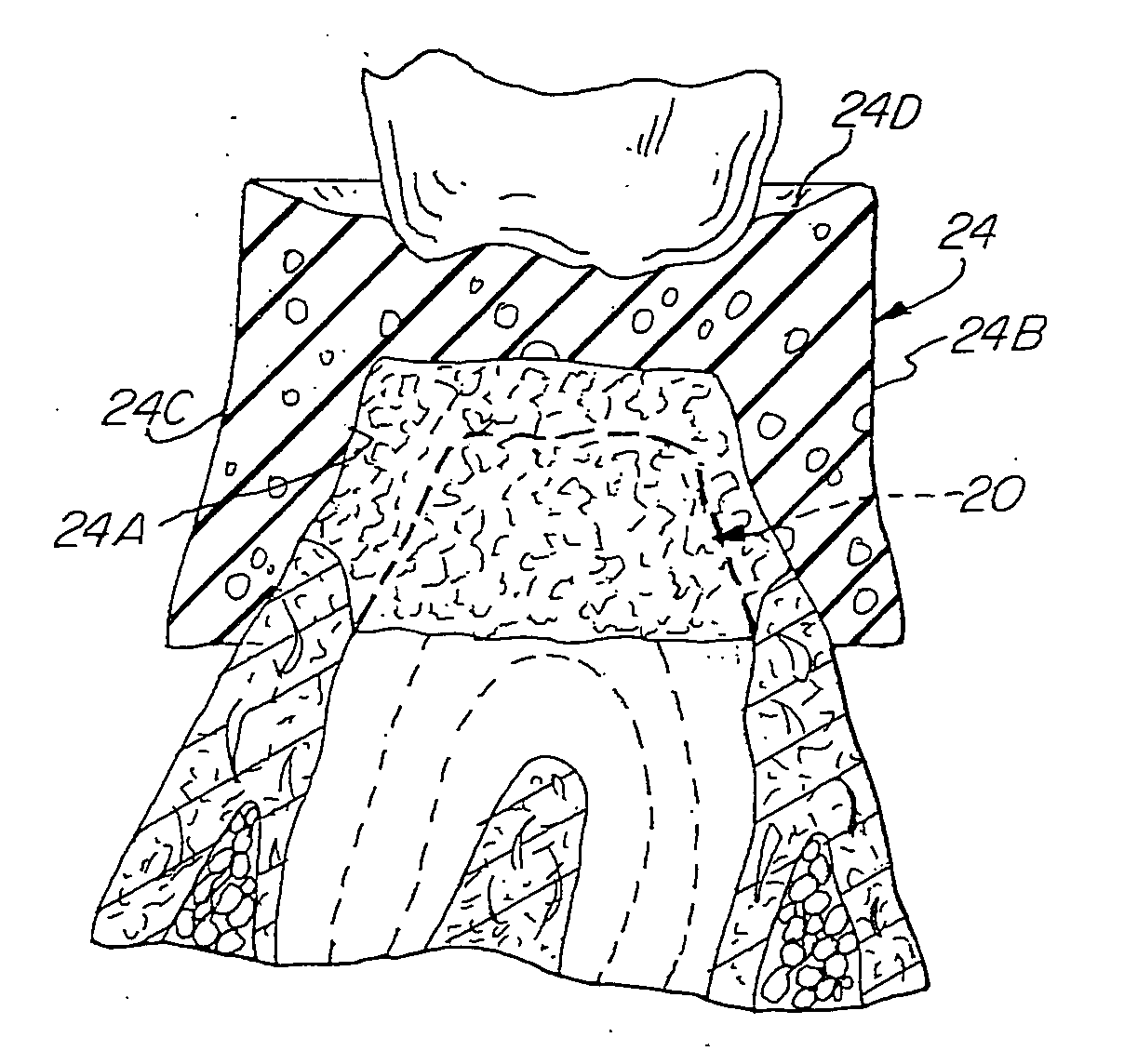
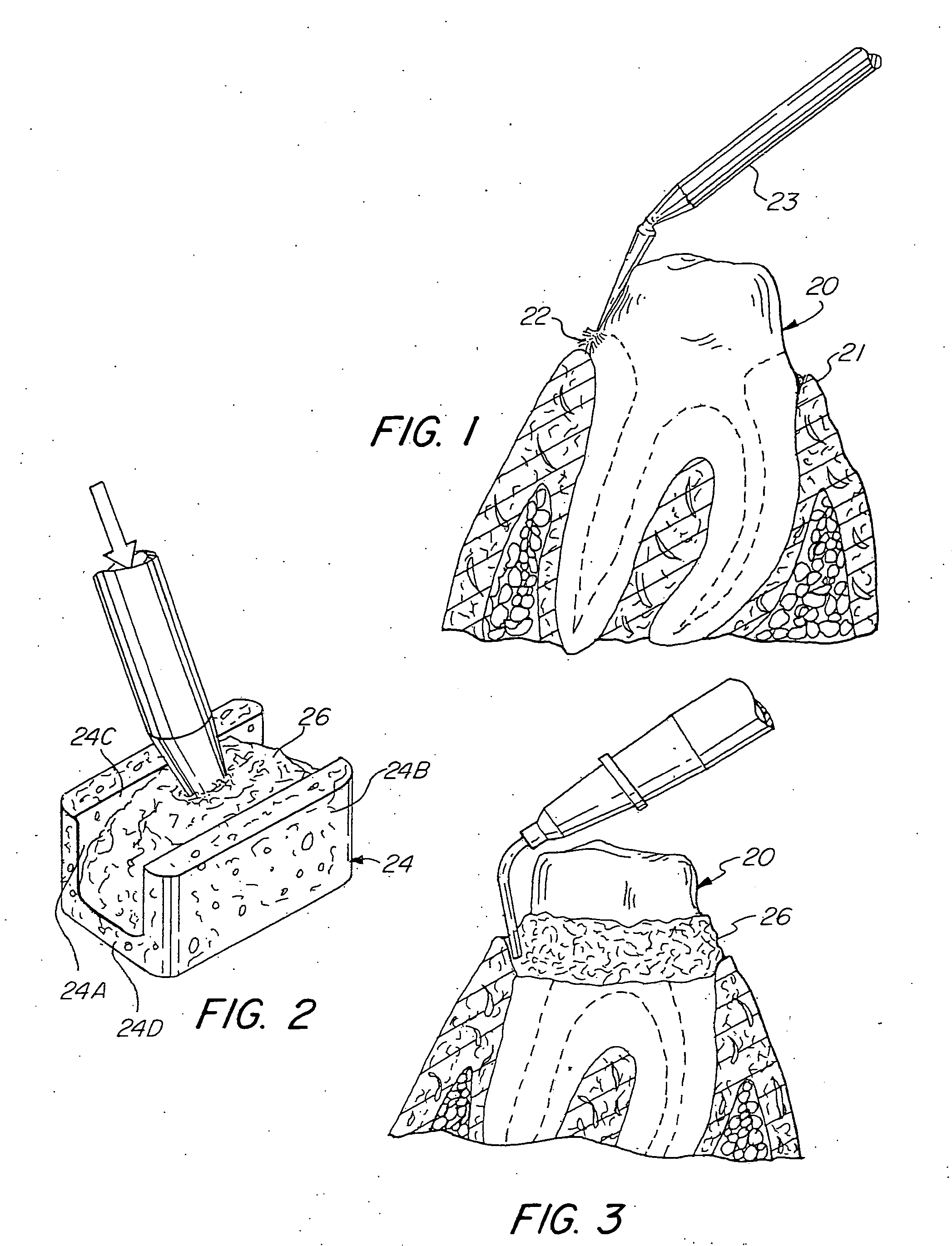
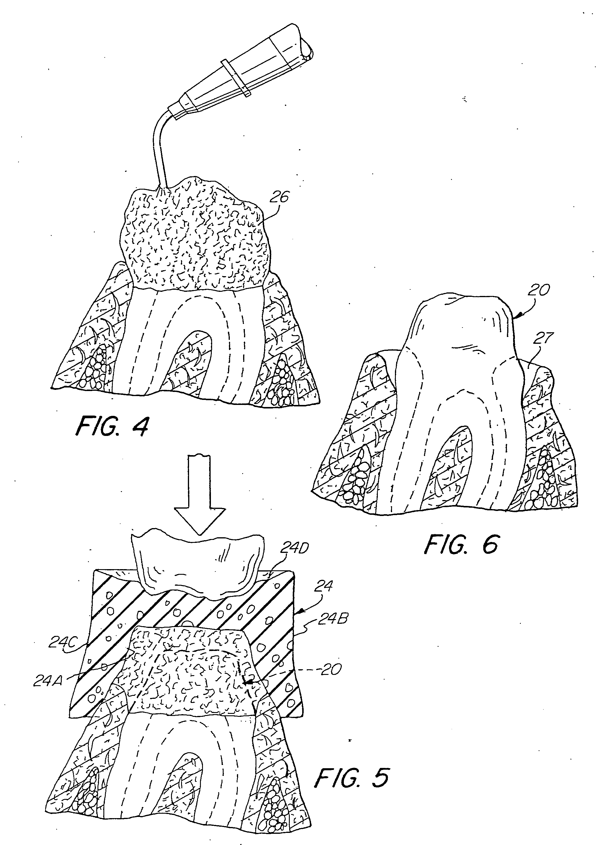
[0021] Referring to the drawings, there is shown a tooth 20 which has been prepared for receiving a crown or bridge. However, before the impression can be taken for preparing the crown or bridge, it is imperative that the gingival sulcus tissue 21 be retracted in order for the dentist to make an accurate impression of the prepared tooth 20. In accordance with this invention and to control any excessive gingival bleeding, an application of a liquid hemostatic agent 22, e.g. aluminum chloride, ferric sulfate or other suitable astringent is applied to the cut tissue in the area of the gingival sulcus. The astringent can be applied with Centrix's Benda micro applicator 23 as seen in FIG. 1, or by any other suitable applicator, e.g. Centrix, Inc.'s BENDA® brush, SoftStix™ disposable applicator, or syringe, and the like. The astringent 22 is applied with moderate pressure and by rubbing the astringent solution against the cut tissue to infuse the astringent solution into the cut capillari...


PUM


Abstract

A method and a device for effecting the cordless retraction of the gingival sulcus tissue prior to the taking of an impression of a tooth for making a crown or bridge which is attained by controlling any bleeding in the gingival sulcus area, and utilizing a dental dam preferably formed of a sponge or foam like material to contain an astringent fortified silicone impression material embedded about the prepared tooth, and using the patient's biting force to apply the necessary pressure onto the dam until the silicone impression material sets and adheres to the dam to enhance easy removal of the set impression material from the tooth. The dam is formed to accommodate either the posterior teeth or the anterior teeth.

Description

RELATED APPLICATIONS [0001] This application is a continuation application of application Ser. No. 10 / 307,695, filed Dec. 2, 2002, which is herein incorporated by reference.FIELD OF THE INVENTION [0002] This invention is directed to a method and device for effecting hemostasis retraction of tissue, and more specifically to a method and device for the retraction and hemostasis of tissue in preparing a tooth prior to the taking of an impression for forming a crown or bridge dental prosthesis. BACKGROUND OF THE INVENTION [0003] The traditional method for controlling hemostasis and retraction of gingival tissue prior to the taking of an impression of a prepared tooth to form a crown or bridge is to mechanically pack a small length of cord saturated with an astringent about the base of the tooth to enlarge the gingival sulcus space about the base of a tooth. After a period of time, the cord is removed from the enlarged space defined about the base of the tooth. Upon the removal of the co...

Claims


Application Information

Patent Timeline
Patent Type & Authority Applications(United States)
IPC IPC(8): A61C9/00
CPCA61C9/0033A61C5/82
Inventor DRAGAN, WILLIAM B.
Owner CENTRIX
Why Eureka
Try Eureka