
Experimental method for promoting tissue engineering pre-vascularization by umbilical cord mesenchymal stem cells
A technology of stem cells and tissue engineering, applied in the medical field, can solve problems such as unclear
Patent Information
- Authority / Receiving Office
- CN · China
- Patent Type
- Applications(China)
- Current Assignee / Owner
- Publication Date
- 2018-03-02
- Estimated Expiration
- Not applicable · inactive patent
Smart Images
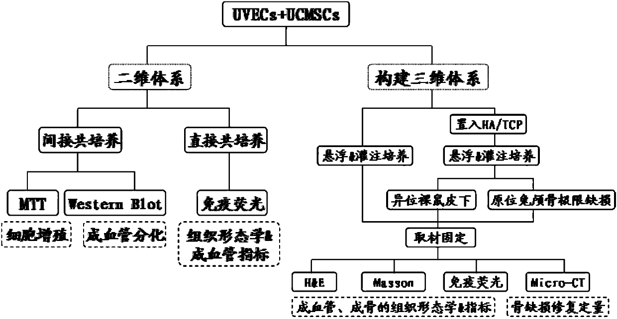
Figure 1 
Figure 2 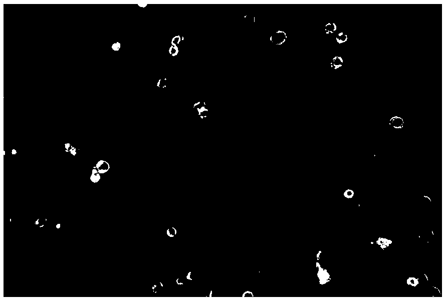
Figure 3
Abstract
Description
technical field
[0001] The invention belongs to the technical field of medicine, and relates to an experimental method for promoting prevascularization of tissue engineering by umbilical cord mesenchymal stem cells. Background technique
[0002] In vitro prevascularization is crucial for successful in vivo transplantation of engineered tissues and organs. Some progress has been made in angiogenesis based on vascular endothelial cells and bioscaffold materials, but how to construct a continuous and stable three-dimensional network microvascular system in vitro is still unclear. Recently, studies have found that umbilical cord mesenchymal stem cells have multilineage differentiation potential and can secrete a variety of cytokines that promote proliferation and differentiation. Our previous work showed that umbilical cord mesenchymal stem cells can participate in and promote the proliferation and angiogenesis differentiation of vascular endothelial cells. Contents of the in...
Examples
Embodiment
[0142] 1. Primary cell culture of umbilical cord mesenchymal stem cells. Such as figure 2 shown.
[0143] 2. Primary culture of umbilical cord vein endothelial cells. Such as image 3 shown.
[0144] 3. Detection of surface markers of umbilical cord mesenchymal stem cells. Such as Figure 4-Figure 10 shown.
[0145] 4. Detection of osteogenic differentiation of umbilical cord mesenchymal stem cells. Such as Figure 11 shown.
[0146] 5. Detection of adipogenic differentiation of umbilical cord mesenchymal stem cells. Such as Figure 12 shown.
[0147] 6. Staining of the colony formation rate of umbilical cord mesenchymal stem cells. Such as Figure 13 shown.
[0148] 7. The umbilical cord vein endothelial cells positively expressed CD-31. Such as Figure 14 shown.
[0149] 8. The umbilical cord vein endothelial cells positively expressed VEGF. Such as Figure 15 shown.
[0150] 9. Umbilical cord vein endothelial cells positively expressed factor Ⅷ. Such as ...