
Use of Novel Chimeric Antigen Receptor Modified T Cells for Cancer Therapy
A chimeric antigen receptor, a new type of technology, applied to genetically modified cells, cells modified by introducing foreign genetic material, receptors/cell surface antigens/cell surface determinants, etc., can solve unsatisfactory curative effects, etc. question
Patent Information
- Authority / Receiving Office
- CN · China
- Patent Type
- Patents(China)
- Current Assignee / Owner
- Publication Date
- 2020-08-18
Smart Images
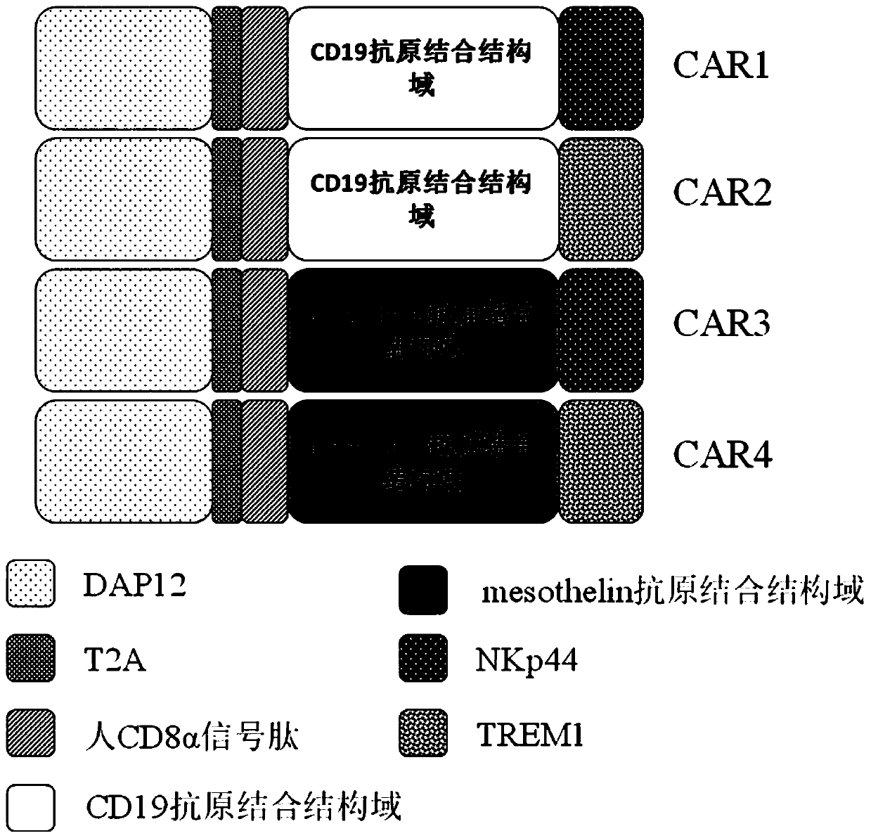
Figure 1 
Figure 2 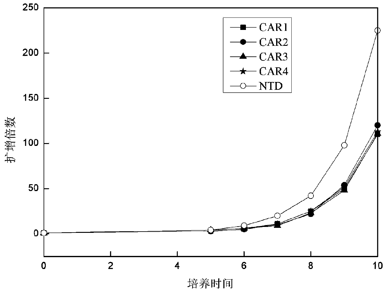
Figure 3
Abstract
Description
technical field
[0001] The invention relates to the technical field of tumor immunotherapy, and provides a composition and a method for treating human cancer. The present invention involves T cells genetically engineered to express a CAR, wherein the CAR includes an antigen binding domain, a first transduction domain and a second transduction domain. The present invention also relates to nucleic acids encoding such transmembrane polypeptides, vectors, and immune cells expressing the CAR on their surface for immunotherapy. The present invention opens the way to an effective adoptive immunotherapy strategy for the treatment of cancer. Background technique
[0002] With the development of tumor immunology theory and clinical technology, chimeric antigen receptor T-cell immunotherapy (CAR-T) has become one of the most promising tumor immunotherapy [Schmitz M, et al . Chimeric antigen receptor-engineered T cells for immunotherapy of Cancer. J Biomed Biotechnol, 2010, doi: 10.11...
Examples
Embodiment 1
[0058] Embodiment 1, chimeric antigen receptor preparation
[0059] The present invention provides a novel chimeric antigen receptor. The chimeric antigen receptor of the present invention is composed of the second intracellular conduction domain-T2A-extracellular signal peptide-antigen binding domain-the first intracellular conduction domain in series. Therefore, viral vectors containing different combinations of stimulation signals need to be constructed separately. In this example, the single-chain antibody targeting CD19 and human mesothelin (mesothelin) are used as the unified extracellular recognition antigen structure, and the following four chimeric antigen receptors need to be constructed respectively ( figure 1 ):
[0060] DAP12-T2A-CD8α signal peptide-CD19 antigen-binding domain-NKp44(CAR1)
[0061] DAP12-T2A-CD8α signal peptide-CD19 antigen-binding domain-TREM1(CAR2)
[0062] DAP12-T2A-CD8α signal peptide-mesothelin antigen-binding domain-NKp44(CAR3)
[0063] ...
Embodiment 2
[0104] Embodiment 2, virus infection T cell
[0105] 1. Isolation and activation of T cells and virus infection
[0106] (1) Isolation of human peripheral blood mononuclear cells
[0107] Use a blood collection tube containing anticoagulant to collect about 10ml of peripheral blood, settle naturally at room temperature (18-25°C) for about 30min, collect the upper layer of plasma, and centrifuge the collected upper layer of plasma at 5000r / min for 10min at a volume ratio of 1:1 Add to the lymphocyte separation medium (purchased from Tianjin Haoyang Biological Products Technology Co., Ltd.), gradient centrifugation, 3000r / min, centrifugation for 30min, after centrifugation, the centrifuge tube is layered from top to bottom: the first layer is the plasma layer ; The second layer is the buffy coat layer of lymphocytes; the third layer is the transparent separation liquid layer; the fourth layer is the red blood cell layer. Aspirate the buffy coat of lymphocytes, wash twice with ...
Embodiment 3
[0112] Example 3. Effect of Virus Infection of CAR-T Cells on Cell Proliferation
[0113]After each group of viruses infected T cells, the T cells were counted every 1-2 days with a complete medium containing 5% autologous plasma + 300 IU / ml recombinant human IL-2 + KBM581. Then observe the growth of T lymphocytes, the results are as follows: image 3 shown. The results showed that after the cells were infected with CAR-expressing viruses, they could still form a typical proliferating clonal group. By counting the cells and drawing the cell proliferation curve, it could be seen that CAR1, CAR2, CAR3, and CAR4 proliferated similarly, compared with T cells not infected with the virus ( image 3 Medium NTD) proliferative ability was slightly weaker.