Liver ultrasonic image identification method based on sparse expression

An ultrasound image and sparse representation technology, which is applied in the field of liver ultrasound image recognition based on sparse representation, and can solve the problems of complex and changeable space-occupying lesions.

Inactive Publication Date: 2016-09-21
SOUTH CHINA UNIV OF TECH
View PDF1 Cites 17 Cited by
  • Summary
  • Abstract
  • Description
  • Claims
  • Application Information

AI Technical Summary

Problems solved by technology

However, due to the inherent shortcomings of liver ultrasound images, such as a large amount of speckle noise and artifacts, and the complexity and changeability of space-occupying lesions

Method used


Image

  • Liver ultrasonic image identification method based on sparse expression
  • Liver ultrasonic image identification method based on sparse expression
  • Liver ultrasonic image identification method based on sparse expression

Examples


Embodiment

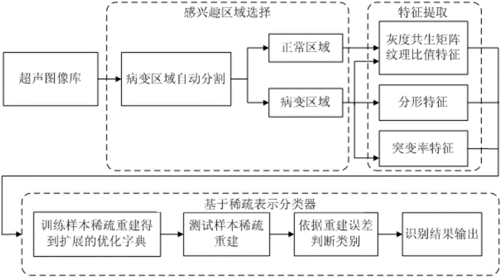
[0068] As shown in Figure 1, a liver ultrasound image recognition method based on sparse representation includes the following steps:

[0069] (1) Select the region of interest from the liver ultrasound image training sample with the space-occupying lesion region, the region of interest includes the space-occupying lesion region R 1 and normal liver area R 2 The liver ultrasound image training samples include liver cyst image samples, hepatic hemangioma image samples, and liver cancer image samples, specifically including the following steps:

[0070] (1-1) Select the space-occupying lesion area R 1 : First, use the region-growing ultrasonic image automatic segmentation algorithm based on energy constraints to outline the edge of the lesion area, then take its circumscribed rectangle, and use the circumscribed rectangle area as the occupying lesion area R 1 ;

[0071] ROI (region of interest) refers to a region selected from the image, which is the focus of image analysis. ...


PUM


Abstract

The invention discloses a liver ultrasonic image identification method based on sparse expression. The method comprises the following steps of: (1) selecting an interested area from a liver ultrasonic image training sample with a space-occupying lesion area; (2) extracting a gray scale symbiosis matrix texture ratio characteristic, a fractal characteristic and a abrupt change rate characteristic of the interested area; (3) utilizing a dictionary expansion method based on sparse reconstruction to construct an expanded dictionary by the image characteristics obtained in the step (2); (4) utilizing the expanded dictionary obtained in the step (3) to construct a classifier based on sparse expression; and (5) inputting the image characteristics of a testing sample into the classifier for identification and judgment, and identifying out the liver ultrasonic image with the space-occupying lesion area. According to the invention, the classified identification accuracy is high, and each index accords with a clinical diagnosis range.

Description

technical field [0001] The invention relates to the technical field of ultrasonic image processing, in particular to a liver ultrasonic image recognition method based on sparse representation. Background technique [0002] Liver cancer ranks sixth in cancer incidence and third in mortality worldwide. Early diagnosis of liver disease is conducive to the early detection and control of liver cancer, improving the survival rate of patients. Ultrasonography has the characteristics of no radiation, simple operation, repeatability, and low cost, so it is widely used in clinical diagnosis of liver diseases. Clinically, the diagnosis of ultrasound images of liver lesions relies on the naked eye observation of doctors to identify them. Not only is the workload huge, but the level of diagnosis depends to a certain extent on the experience of doctors. Therefore, it is of great significance to improve the overall level of ultrasound diagnosis by using medical image processing technolog...

Claims


Application Information

Patent Timeline
IPC IPC(8): G06K9/62G06K9/32G06K9/46
CPCG06V10/25G06V10/40G06V2201/03G06F18/24G06F18/214
Inventor 王伟凝姜怡孜师婷婷
Owner SOUTH CHINA UNIV OF TECH
Why Eureka
Try Eureka