Method for automatically diagnosing cancerous cells

An automatic diagnosis and cell technology, applied in the field of biomedicine, can solve problems such as shortage, and achieve the effect of high discrimination efficiency and high accuracy

Active Publication Date: 2017-10-10
TSINGHUA UNIV
View PDF5 Cites 3 Cited by
  • Summary
  • Abstract
  • Description
  • Claims
  • Application Information

AI Technical Summary

Problems solved by technology

[0003] In related technologies, there is a lack of a means to distinguish cancer cells from healthy cells through computer vision processing methods through the difference in the cell membrane of cancer cells

Method used


Image

  • Method for automatically diagnosing cancerous cells
  • Method for automatically diagnosing cancerous cells

Examples


Embodiment Construction

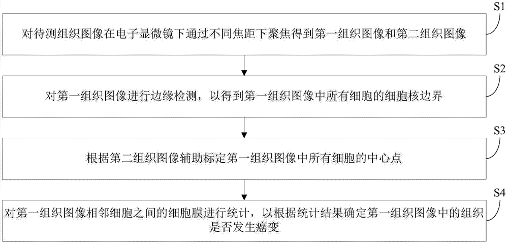
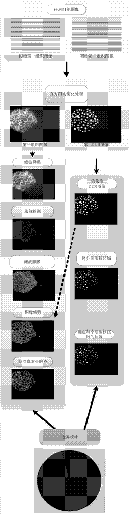
[0017] Embodiments of the present invention are described in detail below, examples of which are shown in the drawings, wherein the same or similar reference numerals designate the same or similar elements or elements having the same or similar functions throughout. The embodiments described below by referring to the figures are exemplary only for explaining the present invention and should not be construed as limiting the present invention.

[0018] In the description of the present invention, it should be understood that the terms "first" and "second" are used for description purposes only, and should not be understood as indicating or implying relative importance.

[0019] These and other aspects of embodiments of the invention will become apparent with reference to the following description and drawings. In these descriptions and drawings, some specific implementations of the embodiments of the present invention are specifically disclosed to represent some ways of implemen...


PUM


Abstract

The present invention discloses a method for automatically diagnosing cancerous cells, comprising: S1: obtaining a first tissue image and a second tissue image by focusing under different electron microscopes through different focal lengths; S2: performing edge detection on the first tissue image to obtain the nucleus boundaries of all the cells in the first tissue image; S3: aiding in the calibration of the center points of all the cells in the first tissue image according to the second tissue image; and S4: statistically counting the cell membranes among adjacent cells of the first tissue image to determine whether canceration occurs in the tissue in the first tissue image or not based on the statistical result. The method of the invention has the following advantages: utilizing the differences existing between cancer cells and healthy cells; and using the computer vision method for automatic characteristics extraction to distinguish the cells at high distinguishing efficiency and accuracy.

Description

technical field [0001] The invention relates to the field of biomedicine, in particular to an automatic diagnosis method for cancerous cells. Background technique [0002] Cancer is a major disease that endangers human health. In clinical medicine, how to diagnose cancer cells is also a very important topic. At this stage, the way of diagnosing cancer tissue is still to manually observe tissue sections, and then make judgments based on experience based on the characteristics of cancer cells. In life sciences, the morphological characteristics of cancer cells are: 1. Changes in cell shape: including cell enlargement, different sizes, and pleomorphism; 2. Changes in the nucleus: including enlarged nuclei, increased ratio of nuclei to plasma, and different sizes of nuclei , abnormal shape, nucleolus hypertrophy, increased number, thickened nuclear membrane and active mitosis; 3, cytoplasmic changes; 4, degeneration and necrosis: cancer cells degeneration and necrosis, cytopla...

Claims


Application Information

Patent Timeline
IPC IPC(8): G06T7/00G06T7/13
CPCG06T7/0012G06T7/13G06T2207/10061G06T2207/30096
Inventor 戴琼海鲍峰索津莉
Owner TSINGHUA UNIV
Why Eureka
Try Eureka